Species ReactivityBovine (Predicted), Dog (Predicted), Human, Mouse, Rat (Predicted)
Immunogen DescriptionA synthetic peptide consisting of amino acids, C-ESKKDTDEVFSS
ConjugateUnconjugated
Purity DescriptionPurified from goat serum by ammonium sulphate precipitation followed by antigen affinity chromatography using the immunizing peptide.
Product DescriptionGoat anti-Nitric oxide synthase 1 (NOS1) Polyclonal Antibody (Unconjugated), suitable for Pep-ELISA, IHC, ICC, FC.
Application(s)FC, ICC, IHC, Pep-ELISA
Application DetailsELISA (1:32000), Western Blot (1-2 µg/mL), Immunohistochemistry (1:3000), Immunofluorescence (10 µg/mL), Flow Cytometry (10 µg/mL). Biosensis recommends optimal dilutions/concentrations should be determined by the end user.
TargetNitric oxide synthase 1 (NOS1)
SpecificityReacts with NOS1 from Human, Mouse.
Target Host SpeciesHuman
Species ReactivityBovine (Predicted), Dog (Predicted), Human, Mouse, Rat (Predicted)
Antibody HostGoat
Antibody TypePolyclonal
Antibody IsotypeIgG
ConjugateUnconjugated
Immunogen DescriptionA synthetic peptide consisting of amino acids, C-ESKKDTDEVFSS
SequenceESKKDTDEVFSS
Purity DescriptionPurified from goat serum by ammonium sulphate precipitation followed by antigen affinity chromatography using the immunizing peptide.
FormatLiquid antibody. Supplied at 0.5 mg/mL in Tris saline, 0.02% sodium azide, pH 7.3 with 0.5% bovine serum albumin.
Storage InstructionsUpon receipt, aliquot and store at -20°C long-term. Store at 2-8°C short-term (up to 2 weeks). Minimize freezing and thawing.
Batch NumberPlease see item label.
Expiration Date12 months after date of receipt (unopened vial).
Scientific BackgroundProduces nitric oxide (NO) which is a messenger molecule with diverse functions throughout the body. In the brain and peripheral nervous system, NO displays many properties of a neurotransmitter. Probably has nitrosylase activity and mediates cysteine S-nitrosylation of cytoplasmic target proteins such SRR (Uniprot).
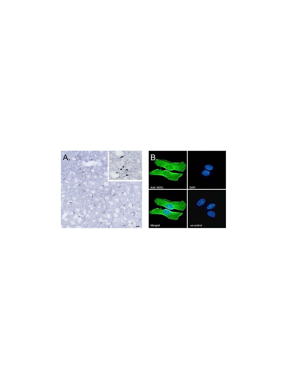
A. G-1870-100 (scale bar: 50 μm; inset: 20 μm) immunostaining of NOS1 neurons in cryosection of a perfusion-fixed (4% PFA) mouse caudate-putamen. HRP-staining with Ni-DAB, after Biotin-SP-anti-goat (IgG) method. Data obtained by Drs. Eva Rumpler and Erik Hrabovszky, Inst, Exp, Med, Budapest, Hungary. B. G-1870-100 Immunofluorescence analysis of paraformaldehyde fixed HeLa cells, permeabilized with 0.15% Triton. Primary incubation 1hr (10 μg/mL) followed by Alexa Fluor 488 secondary antibody (2 μg/mL), showing membrane, cytoplasmic and nuclear staining. The nuclear stain is DAPI (blue). Negative control: Unimmunized goat IgG (10 μg/ml) followed by Alexa Fluor 488 secondary antibody (2 μg/mL).
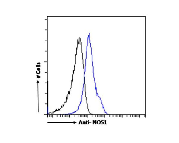
G-1870-100 Flow cytometric analysis of paraformaldehyde fixed Kelly cells (blue line), permeabilized with 0.5%
Triton. Primary incubation 1hr (10 μg/mL) followed by Alexa Fluor 488 secondary antibody (1 μg/mL). IgG control:
Unimmunized goat IgG (lack line) followed by Alexa Fluor 488 secondary antibody.
 1800 605-5127
 +61 (0)8 8352 7711