SpecificityReacts with OGT from Human, Rat. This antibody is expected to recognise both reported isoforms (NP_858058.1 and NP_858059.1.
Species ReactivityBovine (Predicted), Dog (Predicted), Human, Mouse (Predicted), Rat
Immunogen DescriptionA synthetic peptide consisting of amino acids, C-YEHPKDLKLSDGR
ConjugateUnconjugated
Purity DescriptionPurified from goat serum by ammonium sulphate precipitation followed by antigen affinity chromatography using the immunizing peptide.
Product DescriptionGoat anti-UDP-N-acetylglucosamine--peptide N-acetylglucosaminyltransferase 110 kDa subunit (OGT) Polyclonal Antibody (Unconjugated), suitable for Pep-ELISA, WB, IHC, ICC, FC.
Application(s)FC, ICC, IHC, WB, Pep-ELISA
Application DetailsPeptide ELISA (1:64000), Western Blot (0.05-2 µg/mL), Immunohistochemistry (5 µg/mL), Immunofluorescence (10 µg/mL), Flow Cytometry (10 µg/mL). Biosensis recommends optimal dilutions/concentrations should be determined by the end user.
SpecificityReacts with OGT from Human, Rat. This antibody is expected to recognise both reported isoforms (NP_858058.1 and NP_858059.1.
Target Host SpeciesHuman
Species ReactivityBovine (Predicted), Dog (Predicted), Human, Mouse (Predicted), Rat
Antibody HostGoat
Antibody TypePolyclonal
Antibody IsotypeIgG
ConjugateUnconjugated
Immunogen DescriptionA synthetic peptide consisting of amino acids, C-YEHPKDLKLSDGR
SequenceYEHPKDLKLSDGR
Purity DescriptionPurified from goat serum by ammonium sulphate precipitation followed by antigen affinity chromatography using the immunizing peptide.
FormatLiquid antibody. Supplied at 0.5 mg/mL in Tris saline, 0.02% sodium azide, pH 7.3 with 0.5% bovine serum albumin.
Storage InstructionsUpon receipt, aliquot and store at -20°C long-term. Store at 2-8°C short-term (up to 2 weeks). Minimize freezing and thawing.
Batch NumberPlease see item label.
Expiration Date12 months after date of receipt (unopened vial).
Scientific BackgroundGlycosylates a large and diverse number of proteins including histone H2B, AKT1, EZH2, PFKL, KMT2E/MLL5, MAPT/TAU and HCFC1 and can regulate their cellular processes via cross-talk between glycosylation and phosphorylation or by affecting proteolytic processing (Uniprot).
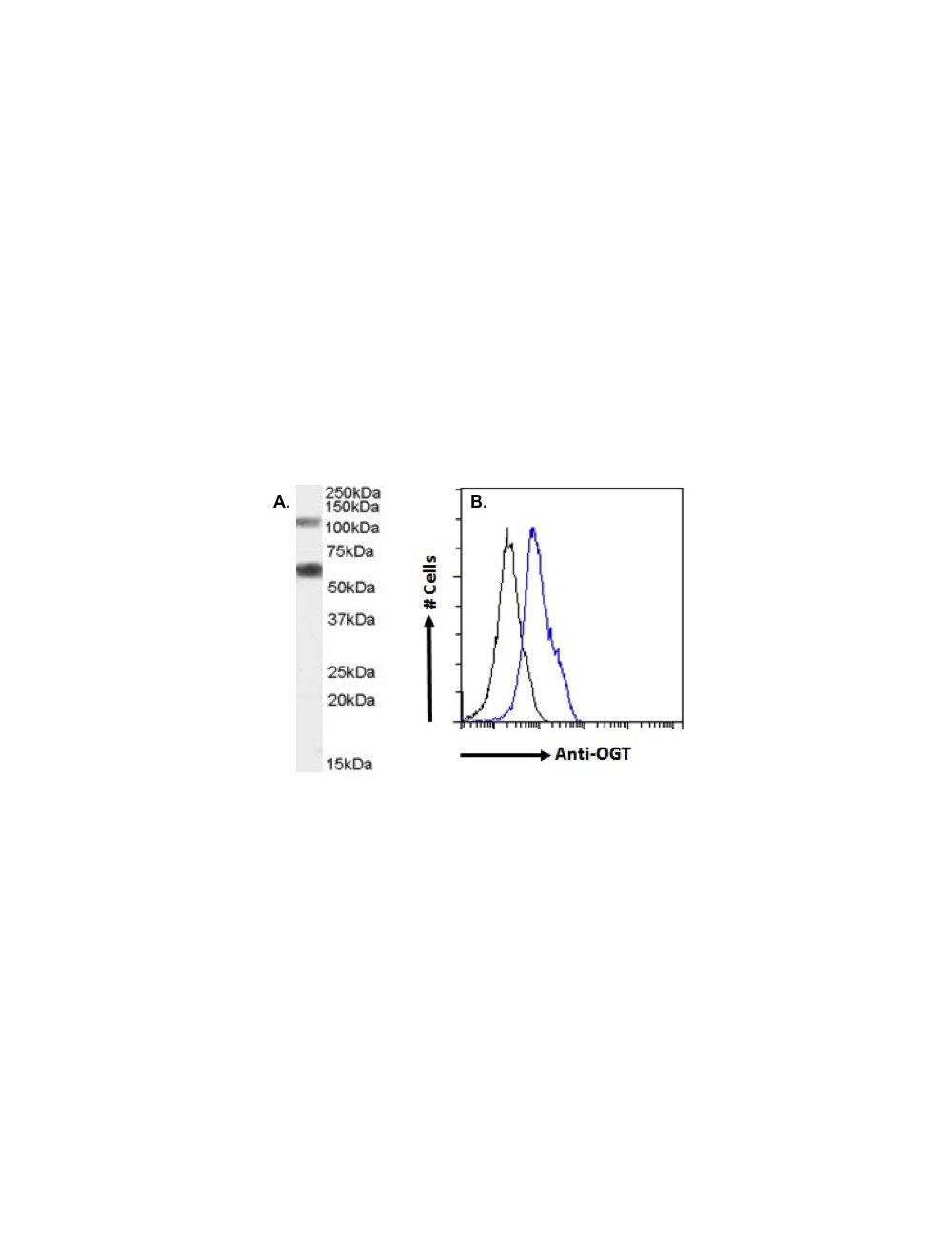
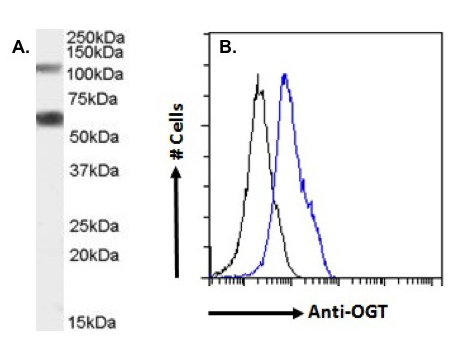
A. Western Blot: (0.05 µg/mL) staining of Rat Pancreas lysate (35 ug protein in RIPA buffer). Detected by chemiluminescence. B. Flow cytometry analysis: of paraformaldehyde fixed HEK293 cells (blue line), permeabilized with 0.5% Triton. Primary incubation 1hr (10 µg/mL) followed by Alexa Fluor 488 secondary antibody (1 µg/mL). IgG control: Unimmunized goat IgG (black line) followed by Alexa Fluor 488 secondary antibody.
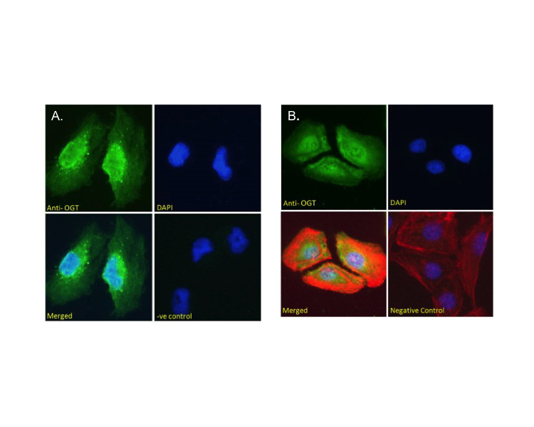
A. Human cortex staining: (5 µg/mL) staining of paraffin embedded Human Cortex. Steamed antigen retrieval with citrate buffer pH 6, AP-staining. B. Immunofluorescence analysis: paraformaldehyde fixed HeLa cells, permeabilized with 0.15% Triton. Primary incubation 1hr (10 µg/mL) followed by Alexa Fluor 488 secondary antibody (2 µg/mL), showing nuclear staining. Actin filaments were stained with phalloidin (red) and the nuclear stain is DAPI (blue). Negative control: Unimmunized goat IgG (10 µg/mL) followed by Alexa Fluor 488 secondary antibody (2 µg/mL).
A. Immunofluorescence analysis: paraformaldehyde fixed U251 cells, permeabilized with 0.15% Triton. Primary incubation 1hr (10 µg/mL) followed by Alexa Fluor 488 secondary antibody (2 µg/mL), showing nuclear and cytoplasmic staining. The nuclear stain is DAPI (blue). Negative control: Unimmunized goat IgG (10 µg/mL) followed by Alexa Fluor 488 secondary antibody (2 µg/mL). B. Immunofluorescence analysis: paraformaldehyde fixed U2OS cells, permeabilized with 0.15% Triton. Primary incubation 1hr (10 µg/mL) followed by Alexa Fluor 488 secondary antibody (2 µg/mL), showing nuclear and membrane/cytoplasmic staining. Actin filaments were stained with phalloidin (red) and the nuclear stain is DAPI (blue). Negative control: Unimmunized goat IgG (10 µg/mL) followed by Alexa Fluor 488 secondary antibody (2 µg/mL).
 1800 605-5127
 +61 (0)8 8352 7711