SpecificityThe specificity of this antibody has been confirmed by WB. This antibody stains bands around 21.5 kDa and 18.5 kDa. A suitable control tissue is rat spinal cord or peripheral nerve homogenate. The major isoforms of MBP run as a closely spaced double of 22 kDa and 18 kDa. Human, Rat
Species ReactivityBovine, Human, Rat
Immunogen DescriptionThree peptide sequences conserved in higher verterbrate MBP protein.
Product DescriptionMouse anti-Myelin basic protein (MBP) Monoclonal Antibody (Unconjugated), suitable for WB, IHC-Frozen, ICC.
Application(s)ICC, IHC-Frozen, WB
Application DetailsWestern Blotting (WB), Immunocytochemistry (ICC), Immunohistochemistry (IHC). IH(P), and Flow Cytometry (~2 ug per10^6 cells). The recommended dilution for WB is 1:5,000-10,0000 and 1:500-1,000 for IC and IH and IH(P). Material should not be over fixed; 2-3 hour post-fixing time is recommended. Long fixations can effect reactivity. In paraffin citrate acid treatment for antigen recovery is recommended. Biosensis recommends optimal dilutions/concentrations should be determined by the end user.
TargetMyelin basic protein (MBP)
SpecificityThe specificity of this antibody has been confirmed by WB. This antibody stains bands around 21.5 kDa and 18.5 kDa. A suitable control tissue is rat spinal cord or peripheral nerve homogenate. The major isoforms of MBP run as a closely spaced double of 22 kDa and 18 kDa. Human, Rat
Target Host SpeciesBovine
Species ReactivityBovine, Human, Rat
Antibody HostMouse
Antibody TypeMonoclonal
Antibody IsotypeIgG
Clone Name7G7
ConjugateUnconjugated
Immunogen DescriptionThree peptide sequences conserved in higher verterbrate MBP protein.
Purity DescriptionProtein G purified
FormatLyophilized from PBS buffer pH 7.2-7.6 with 0.1% trehalose, and sodium azide
Reconstitution InstructionsSpin vial briefly before opening. Reconstitute with 50 µL sterile-filtered, ultrapure water to achieve a 1 mg/mL concentration. Centrifuge to remove any insoluble material.
Storage InstructionsAfter reconstitution of lyophilized antibody, aliquot and store at -20°C for a higher stability. Avoid freeze-thaw cycles.
Batch NumberPlease see item label.
Expiration Date12 months after date of receipt (unopened vial).
Scientific BackgroundMyelin is a membrane characteristic of the nervous tissue and functions as an insulator to increase the velocity of the stimuli being transmitted between a nerve cell body and its target. Myelin isolated from human and bovine nervous tissue is composed of approximately 80% lipid and 20% protein, and 30% of the protein fraction constitutes myelin basic protein (MBP). MBP is an 'intrinsically unstructured' protein with a high proportion (approximately 75%) of random coil, but postulated to have core elements of beta-sheet and alpha-helix. MBP is a major protein in CNS myelin and is expressed specifically in the nervous system. A detailed immunochemical examination of monoclonal and polyclonal antibody responses to MBP and its peptides has revealed the existence of as many as 27 antigenic determinants, many of them conformational. Topological mapping of the potential antigenic determinants onto a model of MBP secondary structure places these determinants within 11 separate regions of the molecule, including those portions that have been found to be encephalitogenic. The message for myelin basic protein is selectively translocated to the ends of the cell processes. Immunization with myelin-associated antigens including MBP significantly promotes recovery after spinal cord contusion injury in the rat model. FUNCTION: Is, with PLP, the most abundant protein component of the myelin membrane in the CNS. Has a role in both the formation and stabilization of this compact multilayer arrangement of bilayers. Each splice variant and charge isomer may have a specialized function in the assembly of an optimized, biochemically functional myelin membrane (By similarity). SUBUNIT: Homodimer (By similarity). SUBCELLULAR LOCATION: Myelin membrane; peripheral membrane protein; cytoplasmic side. Cytoplasmic side of myelin. TISSUE SPECIFICITY: Found in both the central and the peripheral nervous system.
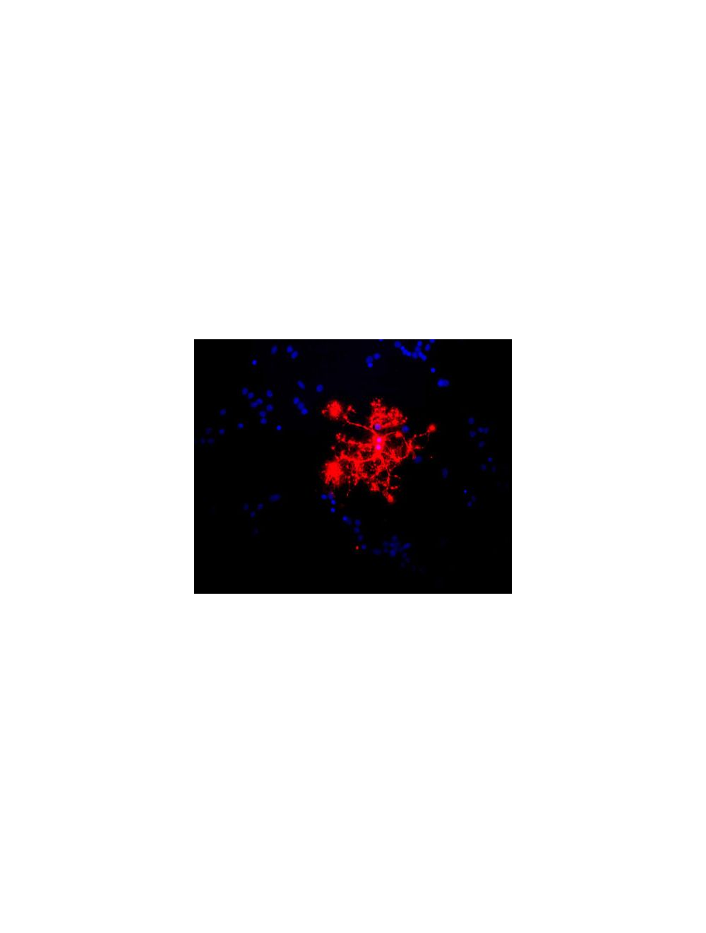
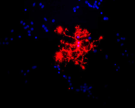
Image shows rat mixed neuron/glial cultures stained with Mouse monoclonal antibody to Myelin Basic Protein [7G7] M-1384-50 (red). Blue is a DNA stain. Note that this antibody stains an oligodendrocyte and some membrane shed from this cell. Other cells in the field include neurons, astrocytes, microglia and fibroblasts, all of which are completely negative.
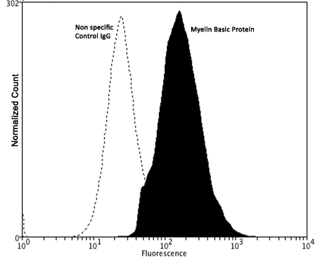
Fixing and Permeabilization of cells: Absolute methanol (10 minutes in ice) and 0.1% Tween-20 in PBS, Blocking: 1% BSA, Primary antibody: Mouse Monoclonal antibody to MBP (cat # M-1384-50, 2 µg per ~10^6 cells) for 30 minutes at room temperature, Secondary antibody: Goat anti-mouse PE labeled secondary antibody (1:100 fold dilution) with incubation for 20 minutes in dark at room temperature. Non-specific Control IgG, clone X63 (cat # M-1249-200) was used as negative control under same conditions (black dashed). Flow cytometry data and results were generated using Orflo MoxiflowTM instrument and protocols. The data demonstrates specific staining of MBP expressed in human neuroblastoma SH-SY5Y cell line using cat # M-1384-50.
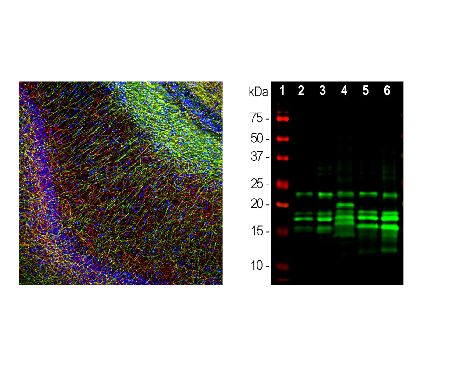
Left: Staining of rat hippocampal section with mouse antibody to myelin basic protein (MBP, 1:5,000, green) by Immunohistochemistry. NF-H was co-stained with R-1389-50 (1:2,000, red). The MBP antibody stains oligodendrocyte cell bodies and the myelin sheathes around axons, while the NF-H antibody labels the axons themselves. Right: Western blot analysis of tissue lysates using mouse antibody to MBP (1:20,000, green): [1] protein standard, [2] rat brain, [3] rat spinal cord, [4] rat sciatic nerve, [5] mouse brain, [6] mouse spinal cord. Multiple bands between 14-22 kDa are alternate transcripts of MBP. Other bands are proteolytic fragments of the MBP protein.
 1800 605-5127
 +61 (0)8 8352 7711