Alternative NamesCoronin-1A; Coronin-like protein A; Clipin-A; Coronin-like protein p57; Tryptophan aspartate-containing coat protein; TACO; CORO1A; CORO1;
Application(s)ICC, WB
Antibody HostRabbit
Antibody TypePolyclonal
SpecificityThe specificity of this antibody has been confirmed by WB and ICC against the antigen. Human, Rat, Mouse and Feline. Predicted to react with other mammalian tissue.
Species ReactivityBovine, Human, Mouse, Pig, Rat
Immunogen DescriptionC-terminal peptide of human coronin 1a coupled to KLH
Application DetailsWestern Blotting (WB) and Immunocytochemistry (ICC). A dilution of 1:2,500-5,000 is recommended for WB. Human Coronin 1A has a predicted length of 461 residues and a MW of 51 kDa. A concentration of 1:500-1:1,000 is recommended for ICC. Biosensis recommends optimal dilutions/concentrations should be determined by the end user.
TargetCoronin- 1A
SpecificityThe specificity of this antibody has been confirmed by WB and ICC against the antigen. Human, Rat, Mouse and Feline. Predicted to react with other mammalian tissue.
Target Host SpeciesHuman
Species ReactivityBovine, Human, Mouse, Pig, Rat
Antibody HostRabbit
Antibody TypePolyclonal
Antibody IsotypeMixed
ConjugateUnconjugated
Immunogen DescriptionC-terminal peptide of human coronin 1a coupled to KLH
Purity DescriptionWhole serum
FormatLyophilized with sodium azide.
Reconstitution InstructionsSpin vial briefly before opening. Reconstitute with 50 µL sterile-filtered, ultrapure water. Centrifuge to remove any insoluble material.
Storage InstructionsAfter reconstitution of lyophilized antibody, aliquot and store at -20°C for a higher stability. Avoid freeze-thaw cycles.
Batch NumberPlease see item label.
Expiration Date12 months after date of receipt (unopened vial).
Alternative NamesCoronin-1A; Coronin-like protein A; Clipin-A; Coronin-like protein p57; Tryptophan aspartate-containing coat protein; TACO; CORO1A; CORO1;
Scientific BackgroundCoronins belong to the WD40 or WD family of proteins. Coronins appear to be particularly involved in binding to actin, actin associated proteins, tubulin and phospholipase C and have been implicated in the mechanisms of chemotaxis and phagocytosis. In mammals there are at least five major coronin proteins, named coronins 1 to 5 in one nomenclature. Another nomenclature divides these five proteins in coronins 1a and 1b, 2a, 2b and 2c (see the Human Genone Organization Gene Nomenclature Committee link for this family). The mammalian coronin family members are abundant components of eukaryotic cells and each type has a restricted cell type specific expression pattern. Coronin 1A is found exclusively in hematopoetic lineage cells such as lymphocytes, macrophages and neutrophils. This antibody is therefore an excellent marker of cells of this lineage and can also be used to study the leading edges particularly of neutrophils. Since the only hematopoetic cells found within the central nervous system are microglia, this antibody is also an excellent marker of this important cell type. Microglia are numerically fairly minor components of the nervous system, but microglial activation is seen in response to a wide variety of damage and disease states, including ALS, Alzheimer's disease and responses to brain tumors. Since coronin 1a is a constitutive component of microglia, the coronin 1a antibody can be used to study both quiescent and activated microglia.
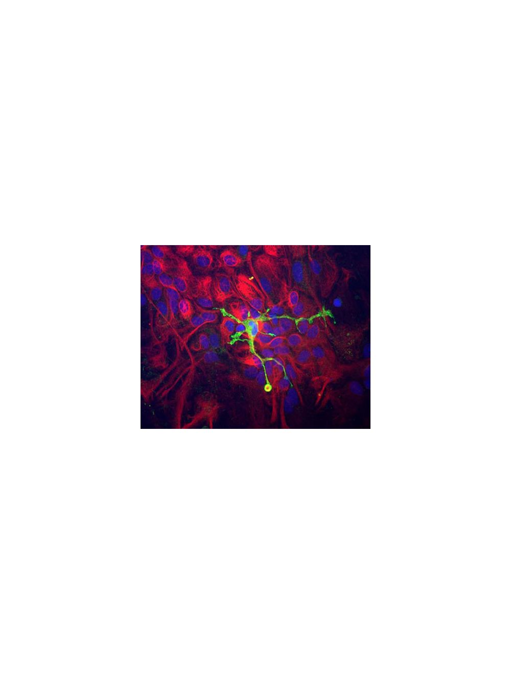
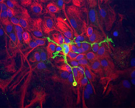
Immunocytochemistry of a mixed neuron/glial culture from newborn rat brain stained with Rabbit polyclonal antibody to Coronin 1a R-1335-50 (green) at a dilution of 1:10,000 and Chicken polyclonal antibody to Vimentin C-1409-50 at 1:5,000. Blue is nuclear DNA counter stain. Glial cells and fibroblasts stain with Vimentin, while microglia alone stain strongly and specifically for Coronin 1a, which can therefore be used as a robust marker of this important cell type.
Left: Detection of Coronin 1a (red) in cortical neuron-glial cell culture from E20 rat by Immunocytochemistry. Coronin 1a antibody was used at 1:1,000 dilution. Cells were co-stained with mouse antibody to GFAP (M-1375-100, green, 1:1,000). Blue: Hoechst nuclear stain. The coronin 1a antibody labels protein expressed in the cytoplasm of microglia cells, while the GFAP antibody stains intermediate filaments in astrocytic cells. Right: Western blot analysis of coronin 1a expression in tissue lysates using rabbit anti-coronin 1a antibody (green, 1:5,000). [1] protein standard, [2] mouse brain, [3] rat brain, [4] cow cerebellum, [5] cow cortex, and [6] pig spinal cord. The strong single band about 55 kDa corresponds to the coronin 1a protein.
Specific ReferencesStephens A.N. et al (2010) Post-translational modifications and protein-specific isoforms in endometriosis revealed by 2D DIGE. J Proteome Res. 2010 May 7;9(5):2438-49. Ahmed Z. et al (2007) Actin-binding proteins coronin-1a and IBA-1 are effective microglial markers for immunohistochemistry. J Histochem Cytochem. 2007 Jul;55(7):687-700.
 1800 605-5127
 +61 (0)8 8352 7711