Product DescriptionRabbit anti-Glial Fibrillary Acidic Protein (GFAP) Polyclonal Antibody (Unconjugated), suitable for WB, ICC, IHC-Frozen.
Application(s)ICC, IHC-Frozen, WB
Application DetailsWestern Blotting (WB), Immunocytochemistry (ICC) and Immunohistochemistry (IHC). A dilution of 1:5,000 is recommended for WB, and 1:1,000 - 1:5,000 for ICC and IHC. A dilution of 1:1,000 using fluorescent secondary antibodies or 1:5,000 using peroxidase or other enzyme-linked methods is recommended for IC. Biosensis recommends optimal dilutions/concentrations should be determined by the end user.
TargetGlial Fibrillary Acidic Protein (GFAP)
Target Host SpeciesHuman
Species ReactivityBovine, Horse, Human, Mouse, Pig, Rat
Antibody HostRabbit
Antibody TypePolyclonal
Antibody IsotypeMixed
ConjugateUnconjugated
Immunogen DescriptionRecombinant full length human GFAP isotype 1 expressed in and purified from E. coli.
Purity DescriptionWhole serum
FormatLyophilized with sodium azide.
Reconstitution InstructionsSpin vial briefly before opening. Reconstitute with 50 µL sterile-filtered, ultrapure water. Centrifuge to remove any insoluble material.
Storage InstructionsAfter reconstitution of lyophilized antibody, aliquot and store at -20°C for a higher stability. Avoid freeze-thaw cycles.
Batch NumberPlease see item label.
Expiration Date12 months after date of receipt (unopened vial).
Alternative NamesAstrocyte; Glial fibrillary acidic protein; GFAP;
Scientific BackgroundGFAP is a 50 kDa intra-cytoplasmic filamentous protein of the cytoskeleton in astrocytes. During the development of the central nervous system, it is a cell-specific marker that distinguishes astrocytes from other glial cells. GFAP immunoreactivity has been shown in immature oligodendrocytes, epiglottic cartilage, pituicytes, papillary meningiomas, myoepithelial cells of the breast and in non-CNS: Schwann cells, salivary gland neoplasms, enteric glia cells, and metastasizing renal carcinomas.
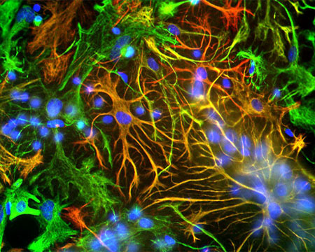
Mixed neuron-glial cultures stained with Rabbit polyclonal antibody to Glial Fibrillary Acidic Protein R-1374-50 (red channel) and Chicken polyclonal antibody to Vimentin C-1409-50 (green channel). The fibroblastic cells contain only Vimentin and so are green, while astrocytes contain either Vimentin and GFAP, so appearing golden, or predominantly GFAP, in which case they appear red. Blue is nuclear DNA stain.
View of a thin section of adult rat cerebellum stained with Chicken polyclonal antibody to Microtubule Associated Protein 2 C-1382-50 (green), Rabbit polyclonal to GFAP R-1374-50 (red) and DNA (blue). The image shows the molecular layer (outside of lobe) and granular layer; blue since its full of small neurons and the white matter in the middle.
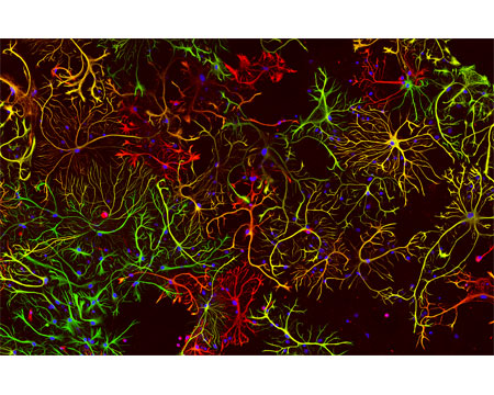
A view of neonatal rat brain cultures stained with Chicken polyclonal antibody to Vimentin C-1409-50 (red) and with Rabbit polyclonal antibody to GFAP R-1374-50 (green). These two proteins are found only in non-neuronal cells so you can\\\\\\\'t see any neurons, except for their nuclei in blue. Maturish astrocytes have only GFAP so appear green or have a mix of both proteins, so appear yellow. Some cells only have the vimentin (immature astrocytes, microglia and fibroblasts) and so appear red.
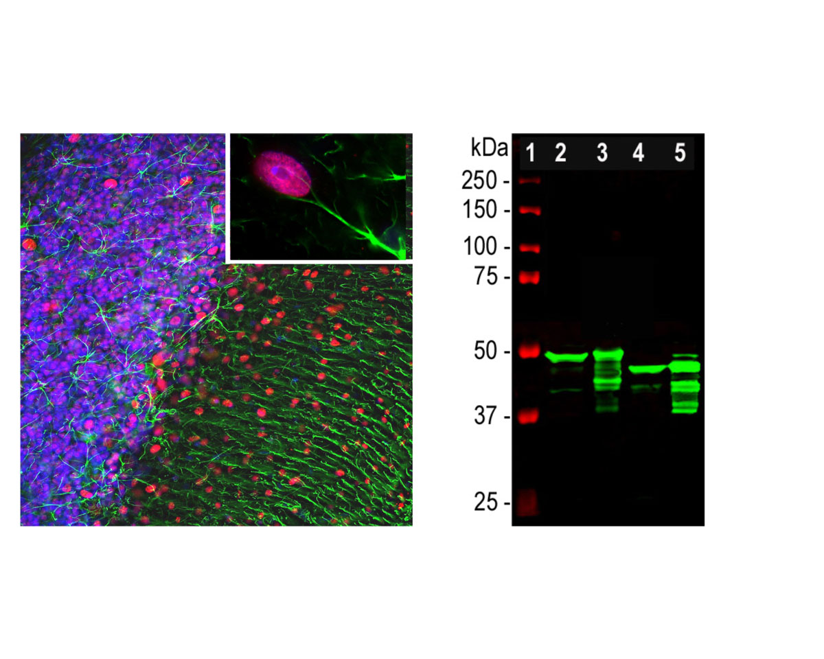
Left: GFAP expression (green) in rat cerebellum section analyzed by Immunohistochemistry. The rabbit anti-GFAP antibody was used at 1:5,000 dilution. Section was co-stained with mouse antibody to MeCP2 (M-1809-100, red, 1:500). Blue: DAPI nuclear stain. IHC Method: Following transcardial perfusion of rat with 4% paraformaldehyde, brain was post-fixed for 1 hour, cut to 45 um, and free-floating sections were stained. The GFAP antibody stains the network of astrocytes and the processes of Bergmann glia in the molecular layer. The MeCP2 antibody specifically labels nuclei of certain neurons. Right: Western blot analysis of tissue lysates using rabbit polyclonal antibody to GFAP (green, 1:5,000). [1] protein standard, [2] rat brain, [3] rat spinal cord, [4] mouse brain, [5] mouse spinal cord. A strong band at about 50 kDa corresponds to the major isotype of the GFAP protein. Smaller isotypes and proteolytic fragments of GFAP are also detected on the blot.
General ReferencesReeves S.A, et al. Proc. Natl. Acad. Sci. U.S.A. 86:5178-5182(1989). Brenner M, et al. Brain Res. Mol. Brain Res. 7:277-286(1990). Isaacs A, et al. Genomics 51:152-154(1998). Ota T, et al. Nat. Genet. 36:40-45(2004). Nielsen A.L, et al. J. Biol. Chem. 277:29983-29991(2002). Singh R, et al. Genomics 82:185-193(2003). Brenner M, et al. Nat. Genet. 27:117-120(2001). Brockmann K, et al. Eur. Neurol. 50:100-105(2003). Stumpf E, et al. Arch. Neurol. 60:1307-1312(2003). Sawaishi Y, et al. Neurology 58:1541-1543(2002). Aoki Y, et al. Neurosci. Lett. 312:71-74(2001).
 1800 605-5127
 +61 (0)8 8352 7711