 1800 605-5127
 +61 (0)8 8352 7711
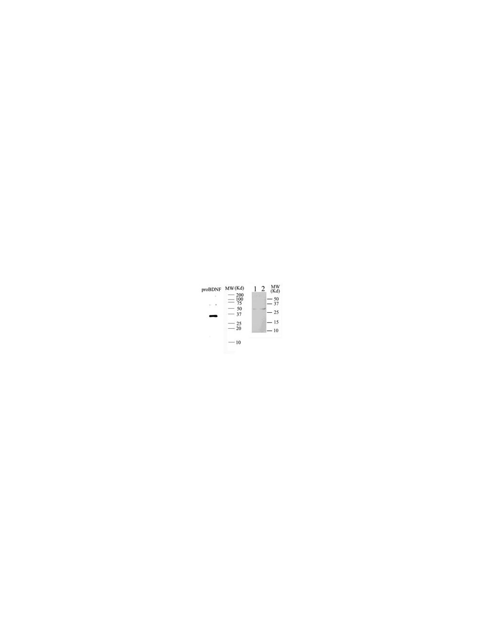

pro-Brain-derived neurotrophic factor (proBDNF), Sheep Polyclonal Antibody
- Product Name pro-Brain-derived neurotrophic factor (proBDNF), Sheep Polyclonal Antibody
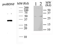
- Product Description Sheep anti-pro-Brain-derived neurotrophic factor (proBDNF) Polyclonal Antibody (Unconjugated), suitable for WB, ELISA, Neutralize.
- Alternative Names Proform brain derived neurotrophic factor
- Application(s) ELISA, Neutralize, WB
- Antibody Host Sheep
- Antibody Type Polyclonal
- Specificity Confirmed to react with purified human proBDNF, crossreact with mouse and rat proBDNF Cross reactivity with other species than human, mouse and rat has not yet been tested
- Species Reactivity Human, Mouse, Rat
- Immunogen Description The recombinant prodomain fragment of human brain-derived neurotrophic factor
- Conjugate Unconjugated
- Purity Description Whole Serum
- Regulatory Status For research use only.
Product Info
- Product Description Sheep anti-pro-Brain-derived neurotrophic factor (proBDNF) Polyclonal Antibody (Unconjugated), suitable for WB, ELISA, Neutralize.
- Application(s) ELISA, Neutralize, WB
- Application Details ELISA, Western Blot, biological neutralization of proBDNF, Immunocytochemistry/Immunofluorescence. Biosensis recommends optimal dilutions/concentrations should be determined by the end user.
- Target pro-Brain-derived neurotrophic factor (proBDNF)
- Specificity Confirmed to react with purified human proBDNF, crossreact with mouse and rat proBDNF Cross reactivity with other species than human, mouse and rat has not yet been tested
- Target Host Species Human
- Species Reactivity Human, Mouse, Rat
- Antibody Host Sheep
- Antibody Type Polyclonal
- Antibody Isotype Mixed
- Conjugate Unconjugated
- Immunogen Description The recombinant prodomain fragment of human brain-derived neurotrophic factor
- Purity Description Whole Serum
- Format Lyophilized
- Reconstitution Instructions Spin vial briefly before opening. Reconstitute in 100 µL sterile-filtered, ultrapure water. Centrifuge to remove any insoluble material.
- Storage Instructions After reconstitution keep aliquots at -20ºC for a higher stability, and at 2-8°C with an appropriate antibacterial agent. Glycerol (1:1) may be added for an additional stability. Avoid repetitive freeze/thaw cycles.
- Batch Number Please see item label.
- Expiration Date 12 months after date of receipt (unopened vial).
- Alternative Names Proform brain derived neurotrophic factor
- Uniprot Number P23560
- Uniprot Number/Name P23560 (BDNF_HUMAN)
- Scientific Background Brain derived neurotrophic factor (BDNF) is synthesized as a precursor (proBDNF) which may be released and have physiological functions to cause cell death. It binds neurotrophin receptor p75 and sortilin and may also be important for the development of nervous system. proBDNF is synthesized in neurons and glia (eg., microglia), transported anterogradely and retrogradely and may be released in an activity dependent manner. This antibody is raised in sheep to detect the prodomain of BDNF and not the mature peptide.
- Shipping Temperature 25°C (ambient)
- UNSPSC CODE 41116161
- Regulatory Status For research use only.
Specifications


-
General References
Fan, YJ. et al (2008) Eur J Neurosci 27(9):2380-90.
Ulman, L. et al (2008) J Neurosci. Oct 29;28(44):11263-8.