Product DescriptiongoogleRabbit anti-Peripherin Polyclonal Antibody (Unconjugated), suitable for WB, ICC.
Alternative NamesPeripherin; Prph; Prph1;
Application(s)ICC, WB
Antibody HostRabbit
Antibody TypePolyclonal
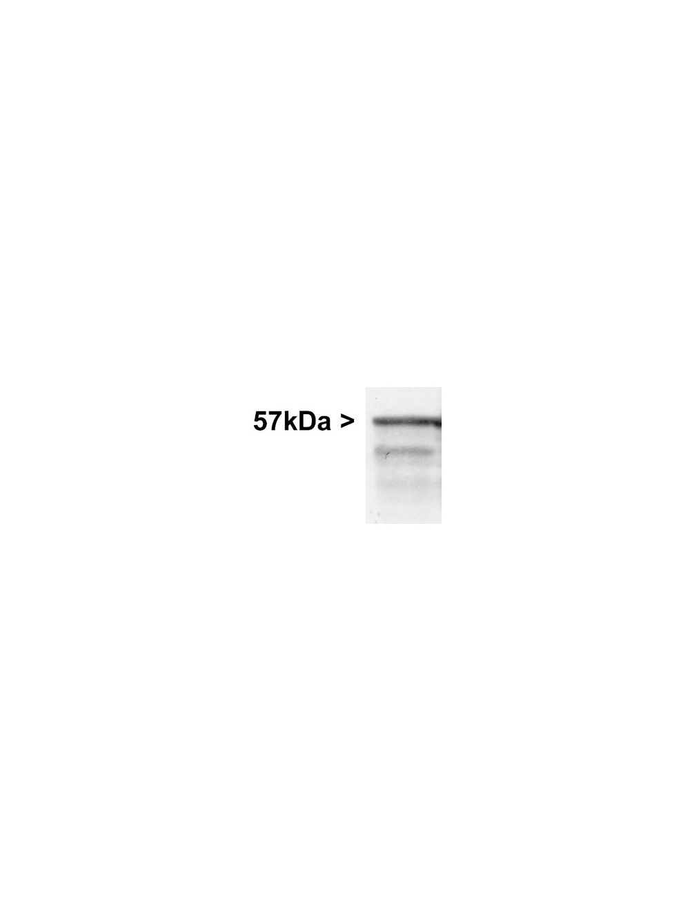
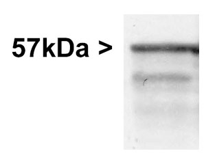
SpecificityThe specificity of this antibody has been confirmed by WB. This antibody detects ~57 kDa Peripherin protein and a smaller molecule derived from Peripherin at ~48 kDa. Hu, Rat, Ms, Fel, Rb. Predicted to react with other mammalian tissue.
Species ReactivityBovine, Human, Mouse, Pig, Rat
Immunogen DescriptionRecombinant full length Peripherin protein expressed in and purified from E.coli.
Product DescriptionRabbit anti-Peripherin Polyclonal Antibody (Unconjugated), suitable for WB, ICC.
Application(s)ICC, WB
Application DetailsWestern Blotting (WB) and Immunocytochemistry (IC). A dilution of 1:2,000 - 1:10,000 is recommended for WB. A dilution of 1:1,00-1:2,000 is recommended for IC. Biosensis recommends optimal dilutions/concentrations should be determined by the end user.
TargetPeripherin
SpecificityThe specificity of this antibody has been confirmed by WB. This antibody detects ~57 kDa Peripherin protein and a smaller molecule derived from Peripherin at ~48 kDa. Hu, Rat, Ms, Fel, Rb. Predicted to react with other mammalian tissue.
Target Host SpeciesHuman
Species ReactivityBovine, Human, Mouse, Pig, Rat
Antibody HostRabbit
Antibody TypePolyclonal
Antibody IsotypeMixed
ConjugateUnconjugated
Immunogen DescriptionRecombinant full length Peripherin protein expressed in and purified from E.coli.
Purity DescriptionWhole serum
FormatLyophilized with sodium azide.
Reconstitution InstructionsSpin vial briefly before opening. Reconstitute with 50 µL sterile-filtered, ultrapure water. Centrifuge to remove any insoluble material.
Storage InstructionsAfter reconstitution of lyophilized antibody, aliquot and store at -20°C for a higher stability. Avoid freeze-thaw cycles.
Batch NumberPlease see item label.
Expiration Date12 months after date of receipt (unopened vial).
Scientific BackgroundPeripherin is a class-III neuronal intermediate filament protein found in certain classes of neuron, most of which are located in the peripheral nervous system.
Western blot of whole rat brain stem homogenate stained with Rabbit polyclonal antibody to Peripherin R-1401-50 at dilution of 1:20,000. A prominent band running with an apparent SDS-PAGE molecular weight of ~57 kDa corresponds to Peripherin. A lower band at ~48 kDa is derived from the Peripherin molecule.
Left: Mixed neuron/glia cultures from newborn rat brain stained with rabbit antibody to peripherin (green) by Immunocytochemistry. Cells were co-stained with chicken antibody to phosphorylated NF-H (C-1386-50, red). A class of large neurons, like the one at the top right of this image, contain peripherin, while the majority of neurons and their processes contain NF-H and not peripherin. Blue: Nuclear stain. Right: Western blot analysis of tissue and cell lysates using rabbit antibody to peripherin (green, 1:10,000). [1] protein standard, [2] rat spinal cord, [3] mouse spinal cord, [4] pig spinal cord, [5] cow spinal cord, [6] SH-SY5Y cells and [7] PC12 cells. The major band at about 57 kDa corresponds to the peripherin protein.
Specific ReferencesIse H. et al (2017) Elucidation of GlcNAc-binding properties of type III intermediate filament proteins, using GlcNAc-bearing polymers. Genes Cell. 2017 Sep; [Epub ahead of print]
Sekerkova G. et al (2008) Espin actin-cytoskeletal proteins are in rat type I spiral ganglion neurons and include splice-isoforms with a functional nuclear localization signal. J Comp Neurol. 2008 Aug 20;509(6):661-76.
 1800 605-5127
 +61 (0)8 8352 7711