1800 605-5127
 +61 (0)8 8352 7711
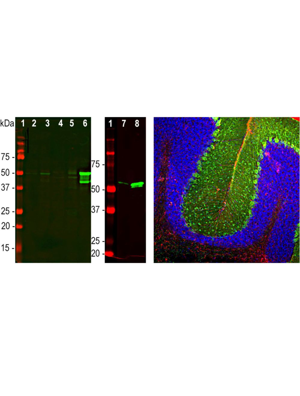

Glial Fibrillary Acidic Protein (GFAP), Mouse Monoclonal Antibody
- Product Name Glial Fibrillary Acidic Protein (GFAP), Mouse Monoclonal Antibody
- Product Description Mouse anti-Glial Fibrillary Acidic Protein (GFAP) Monoclonal Antibody (Unconjugated), suitable for WB, IHC-Frozen.
- Alternative Names Glial fibrillary acidic protein; GFAP
- Application(s) IHC-Frozen, WB
- Antibody Host Mouse
- Antibody Type Monoclonal
- Specificity This antibody is specific for GFAP as demonstrated by western blotting and immunohistochemistry.
- Species Reactivity Bovine, Human, Mouse, Pig, Rat
- Immunogen Description GFAP isolated biochemically from pig spinal cord was used as the immunogen.
- Conjugate Unconjugated
- Purity Description Protein G purified
- Regulatory Status For research use only.
Product Info
- Product Description Mouse anti-Glial Fibrillary Acidic Protein (GFAP) Monoclonal Antibody (Unconjugated), suitable for WB, IHC-Frozen.
- Application(s) IHC-Frozen, WB
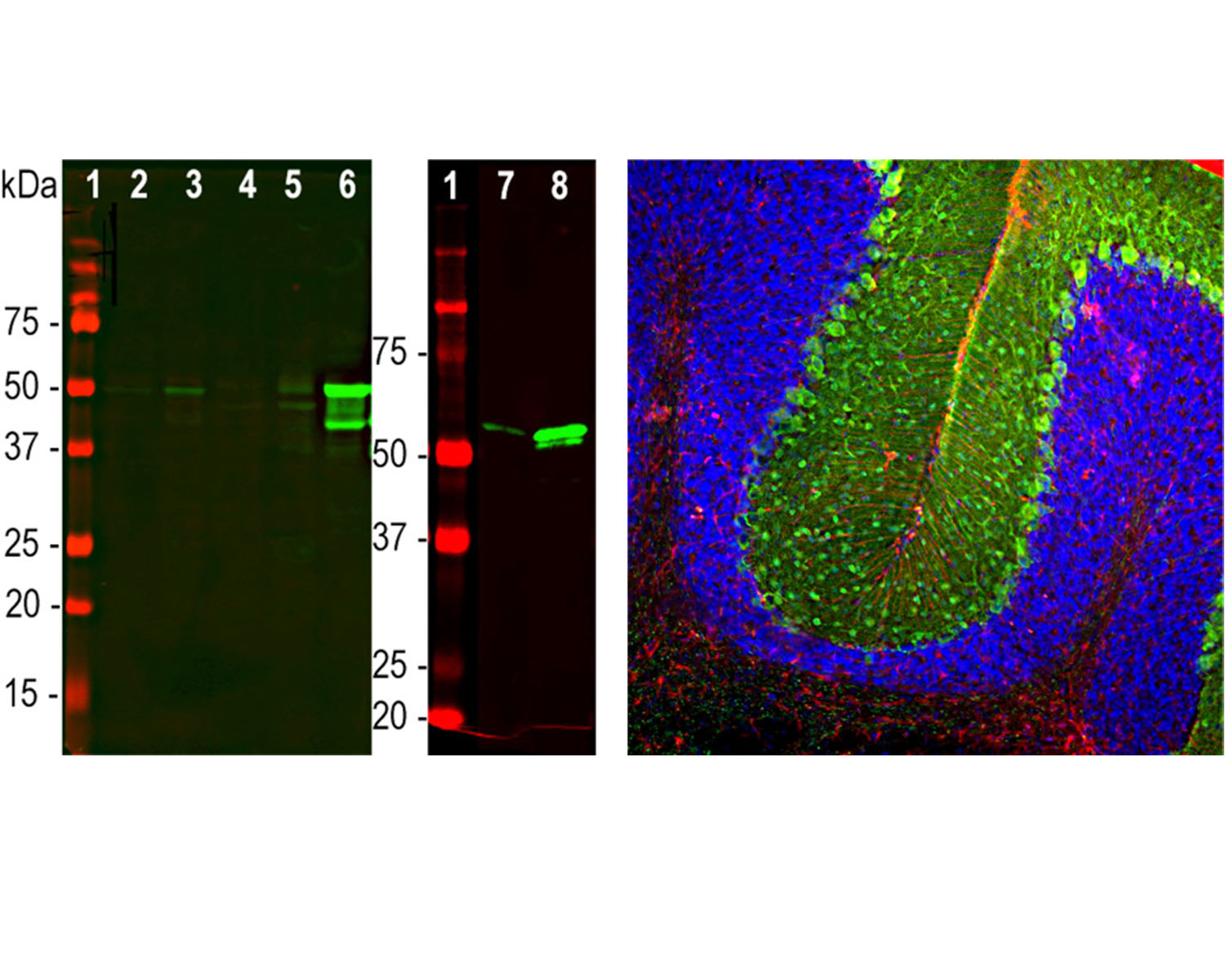
- Application Details Western Blot (1:1,000-1:2,000): tested on rat, mouse brain and spinal cord, human recombinant protein, pig brain. Immunohistochemistry (1:500-1:1,000): tested on rat cerebellum section. Other applications not yet tested. Biosensis recommends optimal dilutions/concentrations should be determined by the end user.
- Target Glial Fibrillary Acidic Protein (GFAP)
- Specificity This antibody is specific for GFAP as demonstrated by western blotting and immunohistochemistry.
- Target Host Species Pig
- Species Reactivity Bovine, Human, Mouse, Pig, Rat
- Antibody Host Mouse
- Antibody Type Monoclonal
- Antibody Isotype IgG1
- Clone Name 2A5
- Conjugate Unconjugated
- Immunogen Description GFAP isolated biochemically from pig spinal cord was used as the immunogen.
- Purity Description Protein G purified
- Format Lyophilized from PBS buffer pH 7.2-7.6 with 0.1% trehalose, and sodium azide
- Reconstitution Instructions Spin vial briefly before opening. Reconstitute with 100 µL sterile-filtered, ultrapure water to achieve a 1 mg/mL concentration. Centrifuge to remove any insoluble material.
- Storage Instructions Store lyophilized antibody at 2-8°C. After reconstitution divide into aliquots and store at -20°C for long-term storage. Store at 2-8°C short-term (up to 4 weeks) with an appropriate antibacterial agent. Avoid repetitive freeze/thaw cycles.
- Batch Number Please see item label.
- Expiration Date 12 months after date of receipt (unopened vial).
- Alternative Names Glial fibrillary acidic protein; GFAP
- Uniprot Number Q864W9
- Uniprot Number/Name Q864W9 (Q864W9_PIG)
- Scientific Background GFAP is a 50 kDa intra-cytoplasmic filamentous protein of the cytoskeleton in astrocytes. During the development of the central nervous system, it is a cell-specific marker that distinguishes astrocytes from other glial cells. GFAP immunoreactivity has been shown in immature oligodendrocytes, epiglottic cartilage, pituicytes, papillary meningiomas, myoepithelial cells of the breast and in non-CNS: Schwann cells, salivary gland neoplasms, enteric glia cells, and metastasizing renal carcinomas.
- Shipping Temperature 25°C (ambient)
- UNSPSC CODE 41116161
- Regulatory Status For research use only.