Product NameGlial fibrillary acidic protein (GFAP), Mouse Monoclonal Antibody
Product DescriptiongoogleMouse anti-Glial fibrillary acidic protein (GFAP) Monoclonal Antibody (Unconjugated), suitable for WB, IHC-Frozen, ICC.
Alternative NamesAstrocyte; Glial fibrillary acidic protein; GFAP;
Application(s)ICC, IHC-Frozen, WB
Antibody HostMouse
Antibody TypeMonoclonal
SpecificityThe specificity of this antibody has been confirmed by WB. Human, Rat, Mouse, Bovine, Porcine. Predicted to react with other mammalian and avian species.
Species ReactivityAvian (Predicted), Bovine, Human, Mouse, Other Mammals (Predicted), Pig, Rat
Immunogen DescriptionPurified GFAP from porcine spinal cord
Product DescriptionMouse anti-Glial fibrillary acidic protein (GFAP) Monoclonal Antibody (Unconjugated), suitable for WB, IHC-Frozen, ICC.
Application(s)ICC, IHC-Frozen, WB
Application DetailsWestern Blotting (WB), Immunocytochemistry (ICC) and Immunohistochemistry (IHC). A dilution of 1:5,000 is recommended for WB. Human GFAP has a predicted length of 432 residues and a MW of 50 kDa. A dilution of 1:500-1:1,000 is recommended for ICC/IHC. This antibody works well on frozen sections, cells in tissue culture and on formalin fixed histological sections. Biosensis recommends optimal dilutions/concentrations should be determined by the end user.
TargetGlial fibrillary acidic protein (GFAP)
SpecificityThe specificity of this antibody has been confirmed by WB. Human, Rat, Mouse, Bovine, Porcine. Predicted to react with other mammalian and avian species.
Target Host SpeciesPig
Species ReactivityAvian (Predicted), Bovine, Human, Mouse, Other Mammals (Predicted), Pig, Rat
Antibody HostMouse
Antibody TypeMonoclonal
Antibody IsotypeIgG1
Clone Name5C10
ConjugateUnconjugated
Immunogen DescriptionPurified GFAP from porcine spinal cord
Purity DescriptionProtein G purified
FormatLyophilized from PBS buffer pH 7.2-7.6 with 0.1% trehalose, and sodium azide
Reconstitution InstructionsSpin vial briefly before opening. Reconstitute with 100 µL sterile-filtered, ultrapure water to achieve a 1 mg/mL concentration. Centrifuge to remove any insoluble material.
Storage InstructionsAfter reconstitution of lyophilized antibody, aliquot and store at -20°C for a higher stability. Avoid freeze-thaw cycles.
Batch NumberPlease see item label.
Expiration Date12 months after date of receipt (unopened vial).
Alternative NamesAstrocyte; Glial fibrillary acidic protein; GFAP;
Scientific BackgroundGFAP is a 50 kDa intra-cytoplasmic filamentous protein of the cytoskeleton in astrocytes. During the development of the central nervous system, it is a cell-specific marker that distinguishes astrocytes from other glial cells. GFAP immunoreactivity has been shown in immature oligodendrocytes, epiglottic cartilage, pituicytes, papillary meningiomas, myoepithelial cells of the breast and in non-CNS: Schwann cells, salivary gland neoplasms, enteric glia cells, and metastasizing renal carcinomas.
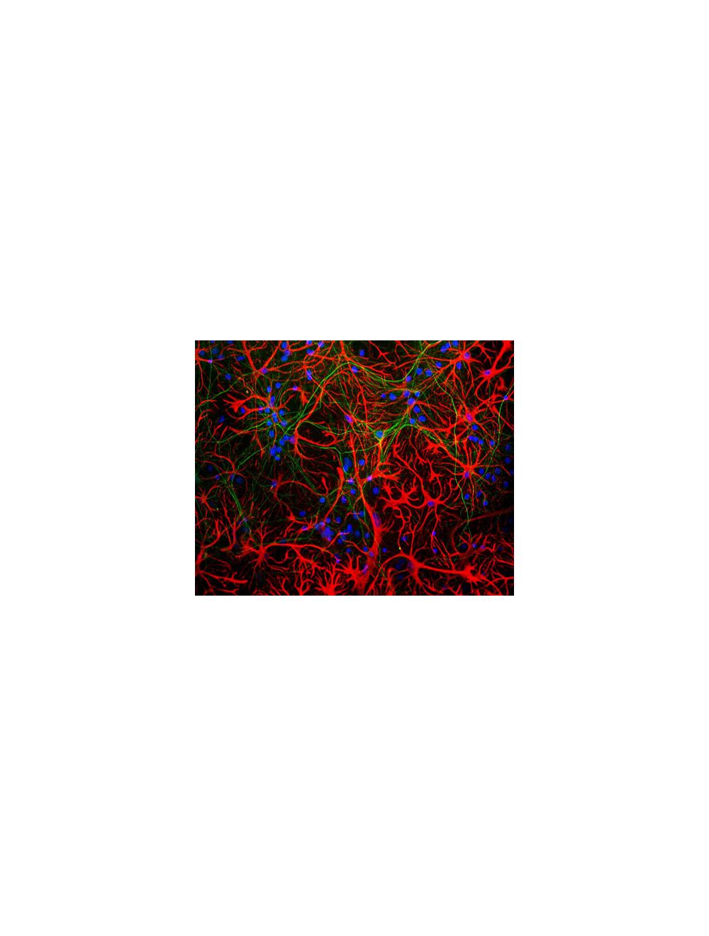
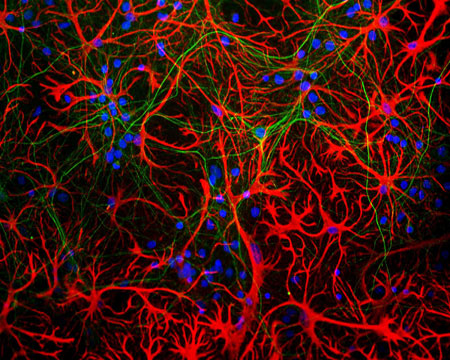
Mixed neuron-glial cultures stained with Mouse monoclonal antibody to Glial Fibrillary Acidic Protein [5C10] M-1375-100 (red) and chicken polylclonal antibody to neurofilament L C-1390-50 (green). The GFAP antibody stains the network of astrocytes in these cultures, while the NF-L antibody stains neurons and their processes. The blue channel shows the localization of DNA.
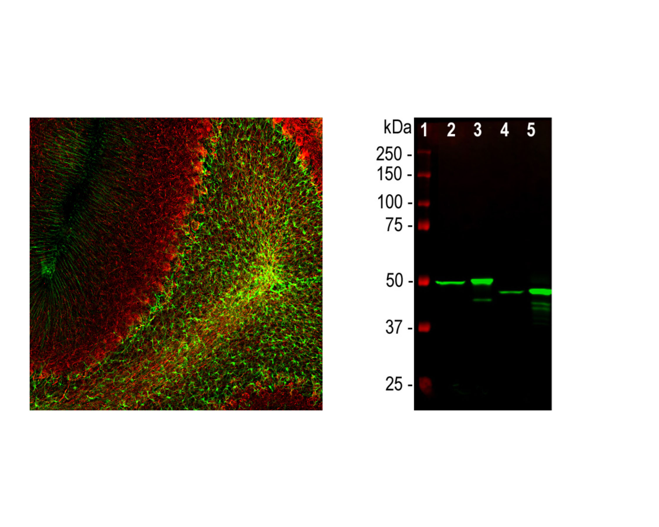
Right: Detection of GFAP expression in rat cerebellum by Immunohistochemistry with mouse antibody to GFAP (1:1,000, green), and co-stained with rabbit antibody to NF-L (R-1392-50, 1:2,000, red). IHC Method: Following transcardial perfusion with 4% PFA, brain was post-fixed for 24 hours, cut to 45 um sections, and free-floating sections were stained. Clone 5C10 stains a network of astroglial cells, while the NF-L antibody labels neuronal cells and their processes. Right: Western blot analysis of whole tissue lysates using mouse antibody to GFAP (1:2,000, green). [1] protein standard , [2] rat brain, [3] rat spinal cord, [4] mouse brain, [5] mouse spinal cord. The strong band at about 50 kDa corresponds to the GFAP protein.
Specific ReferencesKawabe K et al. (2017) Transglutaminases Derived from Astrocytes Accelerate Amyloid _ Aggregation. Neurochem Res. [Epub ahead of print]. Application: ICC (cultured rat astrocytes).
Nagai T et al. (2017) Development of an in situ evaluation system for neural cells using extracellular matrix-modeled gel culture. J Biosci Bioeng. 124(4):430-8. Application: IF (artificial gel matrix).
Kawabe T et al. (2017) Microglia Endocytose Amyloid _ Through the Binding of Transglutaminase 2 and Milk Fat Globule EGF Factor 8 Protein. Neurochem Res. [Epub ahead of print] Application: ICC (cultured astrocytes).
Takano K et al. (2017) Inhibition of Gap Junction Elevates Glutamate Uptake in Cultured Astrocytes. Neurochem Res. [Epub ahead of print] Application: ICC (cultured astrocytes).
General ReferencesReeves S.A, et al. Proc. Natl. Acad. Sci. U.S.A. 86:5178-5182(1989). 2. Brenner M, et al. Brain Res. Mol. Brain Res. 7:277-286(1990). Isaacs A, et al. Genomics 51:152-154(1998). Ota T, et al. Nat. Genet. 36:40-45(2004). Nielsen A.L, et al. J. Biol. Chem. 277:29983-29991(2002). Singh R, et al. Genomics 82:185-193(2003). Brenner M, et al. Nat. Genet. 27:117-120(2001). Brockmann K, et al. Eur. Neurol. 50:100-105(2003). Stumpf E, et al. Arch. Neurol. 60:1307-1312(2003). Sawaishi Y, et al. Neurology 58:1541-1543(2002). Aoki Y, et al. Neurosci. Lett. 312:71-74(2001).
 1800 605-5127
 +61 (0)8 8352 7711