SpecificityThe specificity of this antibody has been confirmed by WB. This antibody detects ~57 kDa Peripherin protein. A suitable control tissue is rat spinal cord or peripheral nerve homogenate. Hu, Rat, Ms, Fel, and other mammals
Species ReactivityCat, Human, Mouse, Other Mammals, Rat
Immunogen DescriptionRecombinant full length rat Peripherin protein expressed in and purified from E.coli
Application DetailsWestern Blotting (WB) and Immunocytochemistry (ICC). A dilution of 1:5,000 - 1:10,000 is recommended for WB. A dilution of 1:500-1,000 is recommended for IC. Biosensis recommends optimal dilutions/concentrations should be determined by the end user.
TargetPeripherin
SpecificityThe specificity of this antibody has been confirmed by WB. This antibody detects ~57 kDa Peripherin protein. A suitable control tissue is rat spinal cord or peripheral nerve homogenate. Hu, Rat, Ms, Fel, and other mammals
Target Host SpeciesRat
Species ReactivityCat, Human, Mouse, Other Mammals, Rat
Antibody HostChicken
Antibody TypePolyclonal
Antibody IsotypeIgY
ConjugateUnconjugated
Immunogen DescriptionRecombinant full length rat Peripherin protein expressed in and purified from E.coli
Purity DescriptionIgY
FormatLyophilized IgY preparation, with sodium azide.
Reconstitution InstructionsSpin vial briefly before opening. Reconstitute with 50 µL sterile-filtered, ultrapure water. Centrifuge to remove any insoluble material.
Storage InstructionsAfter reconstitution of lyophilized antibody, aliquot and store at -20°C for a higher stability. Avoid freeze-thaw cycles.
Batch NumberPlease see item label.
Expiration Date12 months after date of receipt (unopened vial).
Scientific BackgroundPeripherin is a class-III neuronal intermediate filament protein found in certain classes of neuron, most of which are located in the peripheral nervous system.
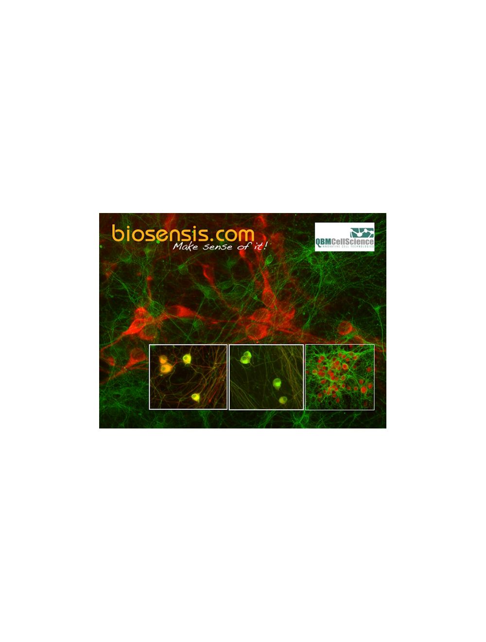
Background image: Substantia Nigra neurons from QBM CellScience thawed and cultured for 5 days and stained with Biosensis rabbit antibody to Tyrosine Hydroxylase (red, Catalog Number R-148-50) and monoclonal antibody Tuj (green). Photo courtesy of QBM Cell Science. Left and middle image: Dorsal Root Ganglia neurons from QBM CellScience thawed and cultured for 7 days and stained with Biosensis chicken antibody to Peripherin (red, Catalog Number C-1399-50) and monoclonal antibody Tuj (green). Photo courtesy of QBM Cell Science. Right image: Substantia Nigra neurons from QBM CellScience thawed and cultured for 14 days and stained with Biosensis rabbit antibody to NeuN (red, Catalog Number R-3770-100) and monoclonal antibody Tuj (green). Photo courtesy of QBM Cell Science.
Image shows rat mixed neuron/glial cultures stained with Chicken polyclonal antibody to Peripherin C-1399-50 (green channel) and Rabbit polyclonal antibody to Internexin alpha R-1379-50 (green channel). These cultures contain mostly neurons which are rich in internexin-alpha, and a subgroup which have a large amount of peripherin also, such as the prominent cell in the middle of the micrograph. Since this cell expresses large amounts of peripherin and internexin-alpha, the green and red signals superimpose to produce a golden cell. Blue is a DNA stain.
Left: Analysis of peripherin expression in mixed fibroblast and PC12 pheochromocytoma cell culture by Immunocytochemistry. Cells were stained with chicken antibody to peripherin (1:5,000, green), and co-stained with rabbit antibody to vimentin (R-1699-100, 1:2,000, red). Blue: DAPI nuclear stain. The peripherin antibody reveals cytoplasmic filamentous staining in the small PC12 cells, while the vimentin antibody stains intermediate filaments in the surrounding fibroblastic cells which do not express peripherin. Right: Western blot analysis of spinal cord tissue lysates (lanes 2-5) and cell lysates (lanes 6 and 7) with chicken antibody to peripherin (1:10,000). [1] protein standard, [2] rat, [3] mouse, [4] pig, [5] cow, [6] SH-SY5Y, [7] PC12. The band at 57 kDa corresponds to peripherin protein.
Specific ReferencesSekerkova G. et al (2008) Espin actin-cytoskeletal proteins are in rat type I spiral ganglion neurons and include splice-isoforms with a functional nuclear localization signal. J Comp Neurol. 2008 Aug 20;509(6):661-76.
 1800 605-5127
 +61 (0)8 8352 7711