SpecificityHuman,reacts with Human, Cow, Rat, Mouse. Antibody is specific for calretinin and does not recognize closely related proteins parvalbumin and calbindin as determined by Western Blotting.
Species ReactivityBovine, Human, Mouse, Rat
Immunogen DescriptionFull-length recombinant human protein
Application DetailsWestern blotting (1:5,000-1:10,000), Immunohistochemistry (1:5,000-1:10,000) and Immunocytochemistry (1:1,000-1:5,000). Biosensis recommends optimal dilutions/concentrations should be determined by the end user.
TargetCalretinin (CR)
SpecificityHuman,reacts with Human, Cow, Rat, Mouse. Antibody is specific for calretinin and does not recognize closely related proteins parvalbumin and calbindin as determined by Western Blotting.
Target Host SpeciesHuman
Species ReactivityBovine, Human, Mouse, Rat
Antibody HostRabbit
Antibody TypePolyclonal
Antibody IsotypeIgG
ConjugateUnconjugated
Immunogen DescriptionFull-length recombinant human protein
Purity DescriptionWhole serum
FormatLyophilized with sodium azide.
Reconstitution InstructionsSpin vial briefly before opening. Reconstitute with 50 µL sterile-filtered, ultrapure water. Centrifuge to remove any insoluble material.
Storage InstructionsStore lyophilized antibody at 2-8°C. After reconstitution divide into aliquots and store at -20°C for long-term storage. Store at 2-8°C short-term (up to 4 weeks) with an appropriate antibacterial agent. Avoid repetitive freeze/thaw cycles.
Batch NumberPlease see item label.
Expiration Date12 months after date of receipt (unopened vial).
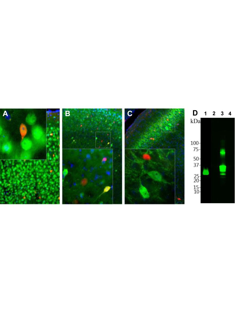
A-C: Immunohistochemisty with rabbit anti-calretinin antibody (red) of adult mouse motor cortex (A), adult mouse visual cortex (B) and adult rat hippocampal CA1 region (C). Fox3/NeuN was co-stained with M-377-100 (green) in motor cortex (A). In visual cortex (B), calbindin was co-labelled with C-1798-50 (green). Double-labelling reveals that calretinin and calbindin antibodies label different population of neurons in the brain. As a result, most cells are labeled with one of the two antibodies and appear to be either red or green. A few cells express both proteins and appear yellow. In rat brain (C), calretinin (red) was co-labelled with mouse antibody to parvalbumin (M-1813-100, green). The two antibodies stain distinct subsets of interneurons in the pyramidal layer, and the positively labeled cells appear to be either red or green. Insets show high magnification pictures of boxed are in each image. Blue: Hoechst-staining of cell nuclei. IHC method: 45 um sections. Tissue was fixed by transcardial perfusion with 4% paraformaldehyde. D: Western blot analysis of calretinin with rabbit anti-calretinin antibody (1:5,000). The antibody recognizes one single band at ~32 kDa in rat brain homogenate (Lane 1, 20 ug total protein loaded). Specificity for calretinin is shown by probing recombinant proteins (0.2 µg/lane) parvalbumin (Lane 2), calretinin (Lane 3) and calbindin (Lane 4).
Left: Rat cerebellum stained with rabbit anti-calretinin (red, 1:5,000) and mouse anti-calbindin (M-1797-100, green, 1:1,000) by Immunohistochemistry. IHC Method: Following transcardial perfusion of rat with 4% paraformaldehyde, brain was post fixed for 24 hours, cut to 45 um, and free-floating sections were stained. The calretinin antibody stains interneurons predominantly in the molecular layer, while the calbindin antibody strongly labels the dendrites and perikarya of Purkinje cells in the molecular layer of the cerebellum. Right: Western blot analysis of [2] rat brain, [3] rat spinal cord, [4] mouse brain, [5] mouse spinal cord, and [6] cow spinal cord (antibody dilution: 1:10,000). A band at 29 kDa corresponds to calretinin protein.
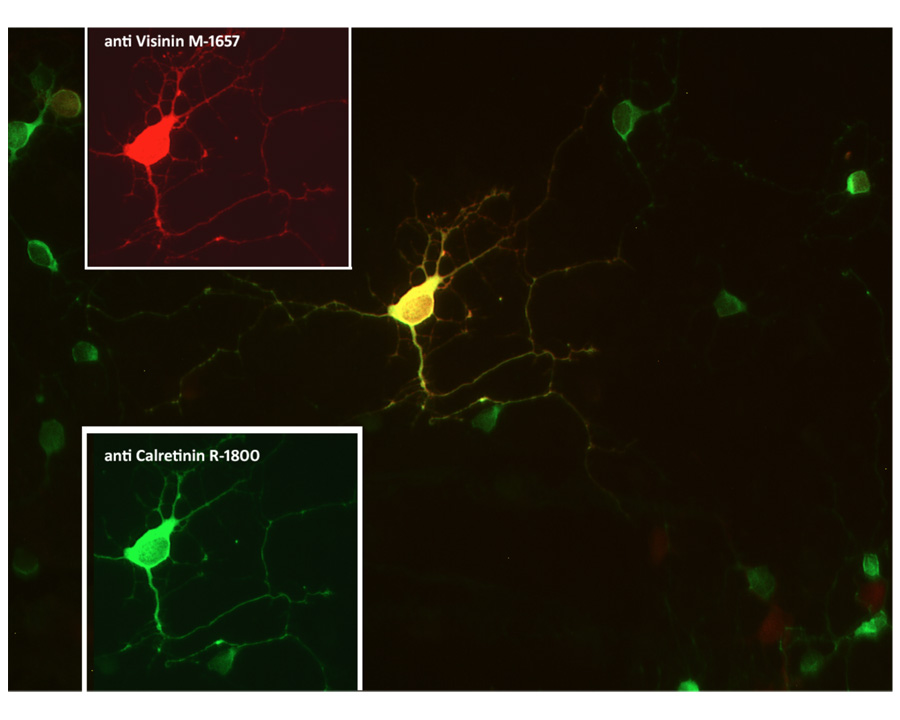
Co-localization of calretinin (green) and visinin (red) in retinal neuron visualized by Immunocytochemistry. Rat retinal primary cells (QBM cat# R-Ret-508) were cultured for 7 days. Image courtesy of QBM Cell Science.
Staining of calretinin (red) and VILIP1 (green) positive neurons by Immunocytochemistry in rat retina culture. Rat retinal primary cells (QBM cat# R-Ret-508) were cultured for 7 days. Image courtesy of QBM Cell Science.
 1800 605-5127
 +61 (0)8 8352 7711