Product NameAdenylate cyclase type III (ACIII), Rabbit Polyclonal Antibody
Product DescriptiongoogleRabbit anti-Adenylate cyclase type III (ACIII) Polyclonal Antibody (Unconjugated), suitable for WB, IHC, ICC.
Application(s)ICC, IHC, WB
Antibody HostRabbit
Antibody TypePolyclonal
SpecificityThe antibody stains a band at about 200 kDa in olfactory epithelium which is rich in cilia. Fewer cilia are found in frontal cortex, and the protein is less heavily glycosylated, and a less prominent band is seen at about 160 kDa. It has also been used successfully for immunocytochemistry on mixed neuron/glia cultures. The antibody has been directly tested for reactivity in rat. It is expected that it will work on human due to homology with the immunogen and possibly other mammal tissues (Human, horse, cow, pig, chicken, mouse).
Species ReactivityHuman (Predicted), Mouse, Other Mammals (Predicted), Rat
Immunogen Description20 amino acid peptide identical to the C-terminus of rat ACIII (amino acids PAAFPNGSSVTLPHQVVDNP)
Product DescriptionRabbit anti-Adenylate cyclase type III (ACIII) Polyclonal Antibody (Unconjugated), suitable for WB, IHC, ICC.
Application(s)ICC, IHC, WB
Application DetailsWestern Blotting (WB), Immunocytochemistry (ICC) and Immunohistochemistry (IHC). A dilution of 1:1,000-1:2,000 is recommended for WB. A dilution of 1:5,000-1:10,000 is recommended for ICC and IHC. Biosensis recommends optimal dilutions/concentrations should be determined by the end user.
TargetAdenylate cyclase type III (ACIII)
SpecificityThe antibody stains a band at about 200 kDa in olfactory epithelium which is rich in cilia. Fewer cilia are found in frontal cortex, and the protein is less heavily glycosylated, and a less prominent band is seen at about 160 kDa. It has also been used successfully for immunocytochemistry on mixed neuron/glia cultures. The antibody has been directly tested for reactivity in rat. It is expected that it will work on human due to homology with the immunogen and possibly other mammal tissues (Human, horse, cow, pig, chicken, mouse).
Target Host SpeciesRat
Species ReactivityHuman (Predicted), Mouse, Other Mammals (Predicted), Rat
Antibody HostRabbit
Antibody TypePolyclonal
Antibody IsotypeIgG
ConjugateUnconjugated
Immunogen Description20 amino acid peptide identical to the C-terminus of rat ACIII (amino acids PAAFPNGSSVTLPHQVVDNP)
Purity DescriptionAffinity purified
FormatLyophilized from PBS buffer pH 7.2-7.6 with 0.1% trehalose, and sodium azide
Reconstitution InstructionsSpin vial briefly before opening. Reconstitute with 100 µL sterile-filtered, ultrapure water. Centrifuge to remove any insoluble material.
Storage InstructionsStore lyophilized, unopened vial at 2-8°C or lower. After reconstitution, prepare aliquots and store at -20°C to -80°C for a higher stability. Avoid freeze-thaw cycles.
Batch NumberPlease see item label.
Expiration Date12 months after date of receipt (unopened vial).
Scientific BackgroundAdenylate cyclases are enzymes which interact with and are activated by the GTP bound alpha subunits of trimeric G-proteins. Activated adenylate cyclases are responsible for the production of the important "second messenger" signalling molecule cyclic-AMP, which is generated from ATP. The type III adenylate cyclase enzyme is localized in the membranes surrounding the cilia in neurons, and our antibody is an excellent marker of neuronal cilia in the brain and in cells in tissue culture. Adenylate cyclase type III is a large complex molecule of, in the human, 1145 amino acids with a deduced molecular weight of 129 kDa. The protein may be variably glycosylated, so that on SDS-PAGE and western blots it runs as a diffuse band of about 160 kDa in cortex and about 200 kDa in olfactory epithelium. The molecule has a complex structure, with 12 transmembrane domains and two cyclase domains. Each cyclase domain is immediately C-terminal to 6 transmembrane segments, but only the second, C-terminal cyclase is believed to be catalytically active.
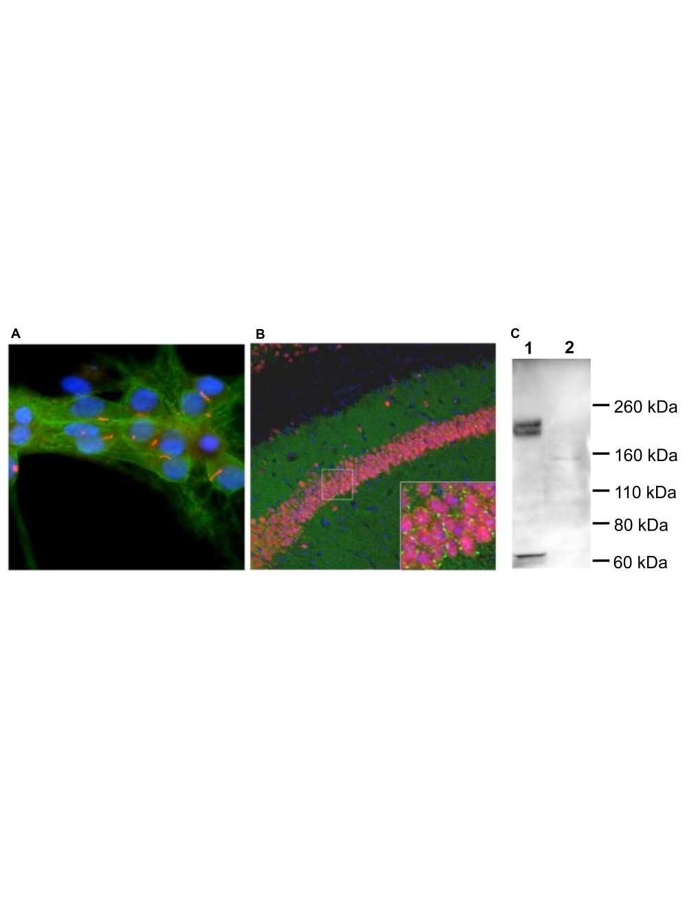
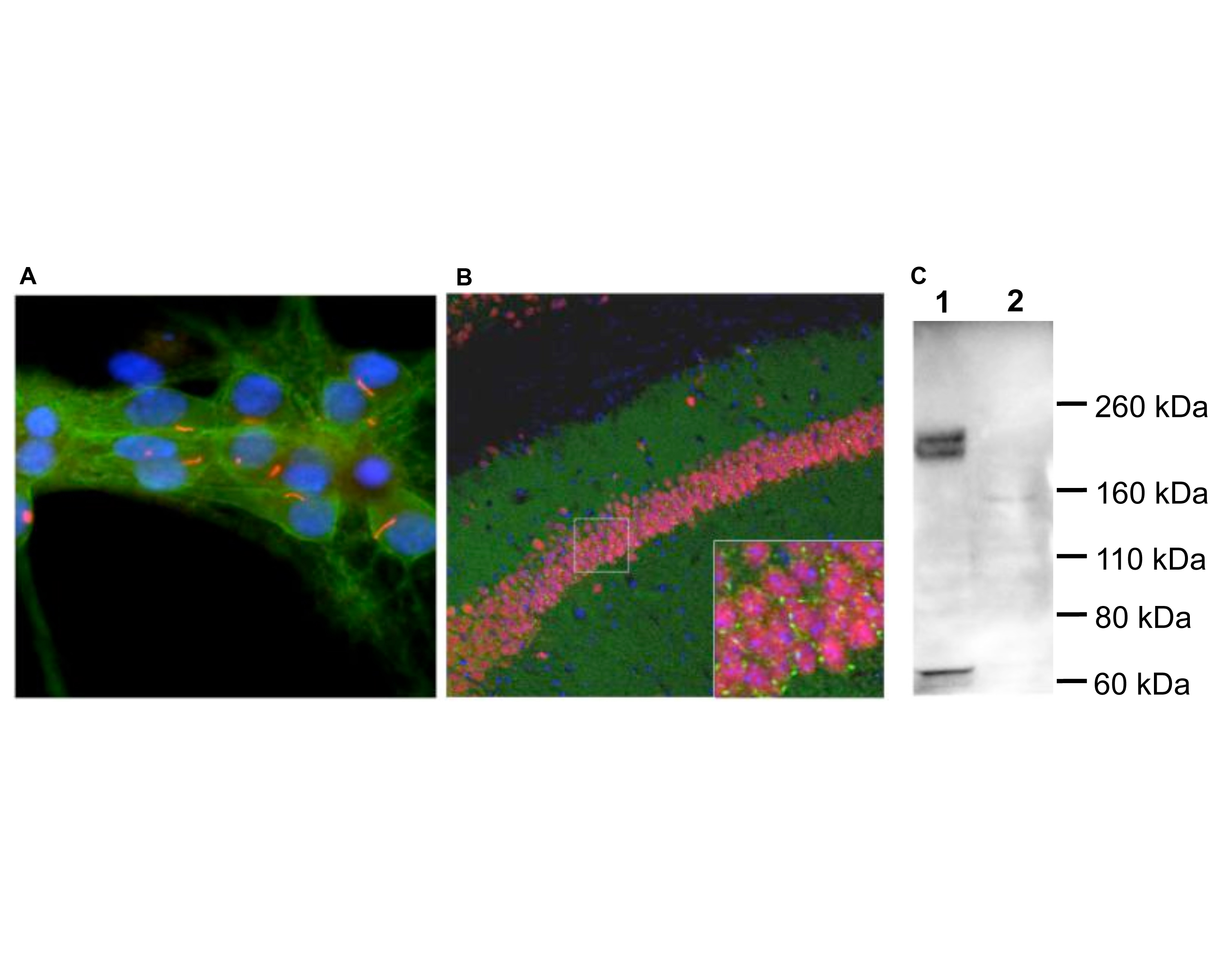
A: Immunofluorescence staining of rat neuron-glial cultures with R-1687-100 (red). The rabbit antibody shows strong and specific staining of neuronal cilia. Neuronal alpha-II spectrin was double-labelled with M-1575-100 (green), and cell nuclei were visualized with a DNA dye (blue). B: Immunohistochemistry of mouse brain sections with R-1687-100 (green). Brains were fixed by transcardial perfusion with 4% paraformaldehyde and stained with R-1687-100 antibody (green) and anti-Fox3/NeuN monoclonal antibody M-377-100 antibody (red). R-1687-100 specifically labels cilia next to the pyramidal neurons in CA1 hippocampus region, but not in other part of the brain. Cell nuclei were stained with DAPI (blue). C: Western blot analysis of rat olfactory epithelium (Lane 1) and frontal cortex (Lane 2). R-1687-100 reveals protein bands at about 200 kDa in olfactory epithelium, a tissue which is rich in cilia. The frontal cortex contains less cilia, and the protein is also less heavily glycosylated, so that a much less prominent band is seen at about 160 kDa.
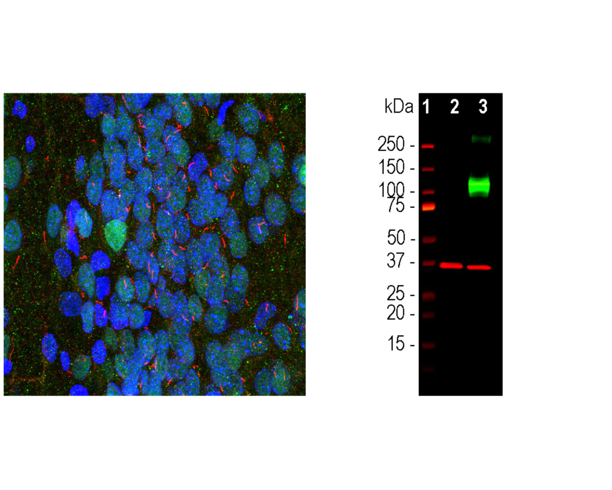
Left: Analysis of rat hippocampus with rabbit antibody to adenylate cyclase III (red) by Immunocytochemistry. Section was co-stained for MeCP2 (green). Blue: DAPI nuclear stain. The ACIII antibody reveals neuronal cilia, while the MeCP2 antibody reveals the neuronal nuclei. Right: Western blot analysis of HEK293 cell lysates using rabbit antibody to ACIII (green, 1:2,000). [1] protein standard, [2] non-transfected HEK293 cells, and [3] HEK293 cells transfected with an expression construct containing a Myc-DDK tagged full length human adenylate cyclase III cDNA. The strong band at about 130 kDa in the transfected cells demonstrates overexpression of the human ACIII protein, and the bands over 250 kDa presumably correspond to either glycosylated or aggregated forms of ACIII. The same blot was simultaneously probed with mouse antibody to GAPDH (M-1376-250, red, 1:5,000, lanes 2-3), which reveals a single band at about 37 kDa in both transfected and non-transfected cells.
General ReferencesFuchs JL, Schwark HD. Neuronal primary cilia: a review. Cell Biol Int. 28:111-8 (2004). Louvi A and Grove EA. Cilia in the CNS: the quiet organelle claims center stage. Neuron 69:1046-1060 (2011).
 1800 605-5127
 +61 (0)8 8352 7711