View of a thin section of adult rat cerebellum stained with Chicken polyclonal antibody to Microtubule Associated Protein 2 C-1382-50 (green), Rabbit polyclonal to GFAP R-1374-50 (red) and DNA (blue). The image shows the molecular layer (outside of lobe) and granular layer; blue since its full of small neurons and the white matter in the middle.
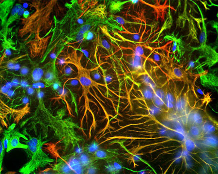
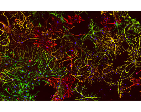
A view of neonatal rat brain cultures stained with Chicken polyclonal antibody to Vimentin C-1409-50 (red) and with Rabbit polyclonal antibody to GFAP R-1374-50 (green). These two proteins are found only in non-neuronal cells so you can\\\\\\\'t see any neurons, except for their nuclei in blue. Maturish astrocytes have only GFAP so appear green or have a mix of both proteins, so appear yellow. Some cells only have the vimentin (immature astrocytes, microglia and fibroblasts) and so appear red.
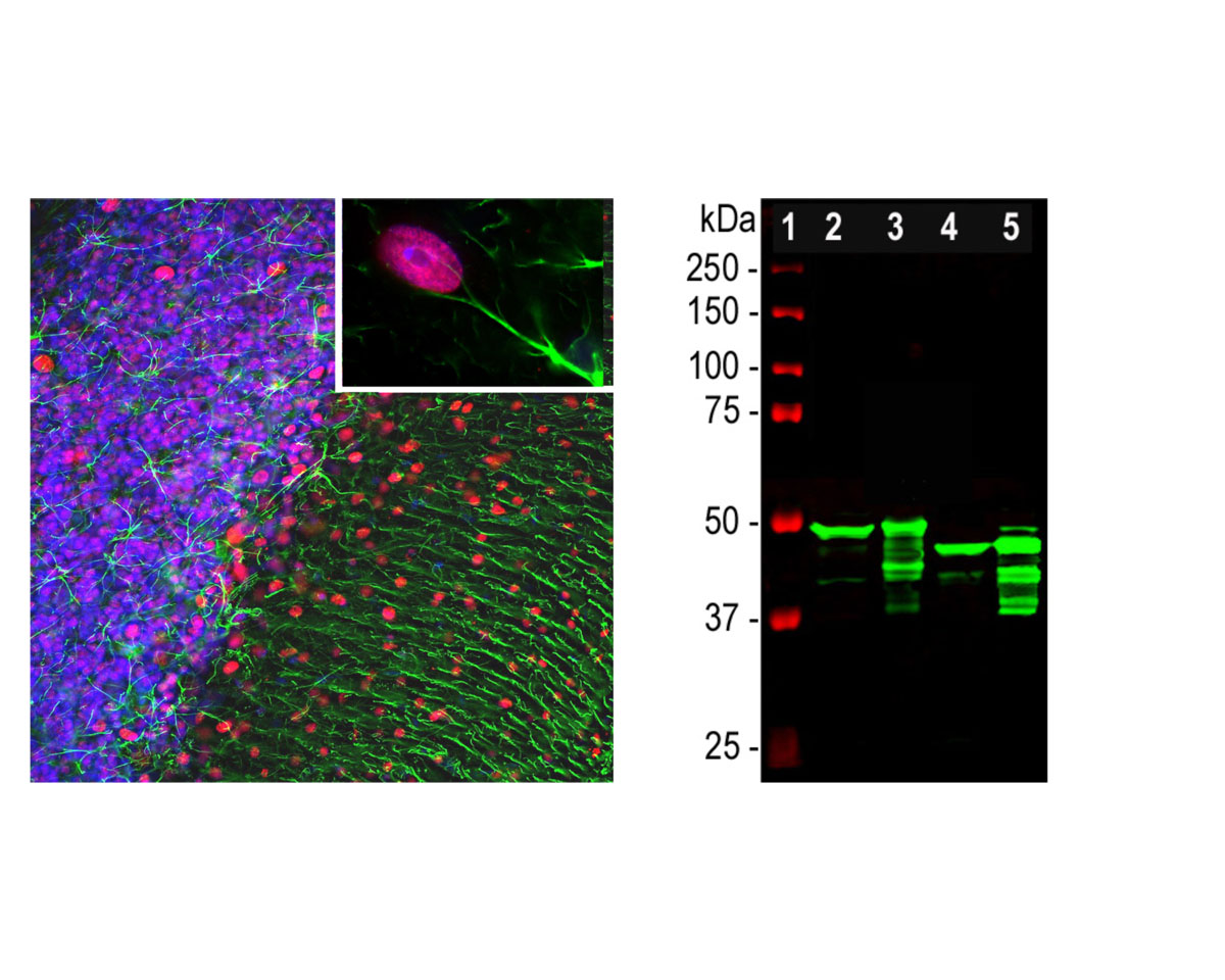
Left: GFAP expression (green) in rat cerebellum section analyzed by Immunohistochemistry. The rabbit anti-GFAP antibody was used at 1:5,000 dilution. Section was co-stained with mouse antibody to MeCP2 (M-1809-100, red, 1:500). Blue: DAPI nuclear stain. IHC Method: Following transcardial perfusion of rat with 4% paraformaldehyde, brain was post-fixed for 1 hour, cut to 45 um, and free-floating sections were stained. The GFAP antibody stains the network of astrocytes and the processes of Bergmann glia in the molecular layer. The MeCP2 antibody specifically labels nuclei of certain neurons. Right: Western blot analysis of tissue lysates using rabbit polyclonal antibody to GFAP (green, 1:5,000). [1] protein standard, [2] rat brain, [3] rat spinal cord, [4] mouse brain, [5] mouse spinal cord. A strong band at about 50 kDa corresponds to the major isotype of the GFAP protein. Smaller isotypes and proteolytic fragments of GFAP are also detected on the blot.
 1800 605-5127
 +61 (0)8 8352 7711