 1800 605-5127
 +61 (0)8 8352 7711
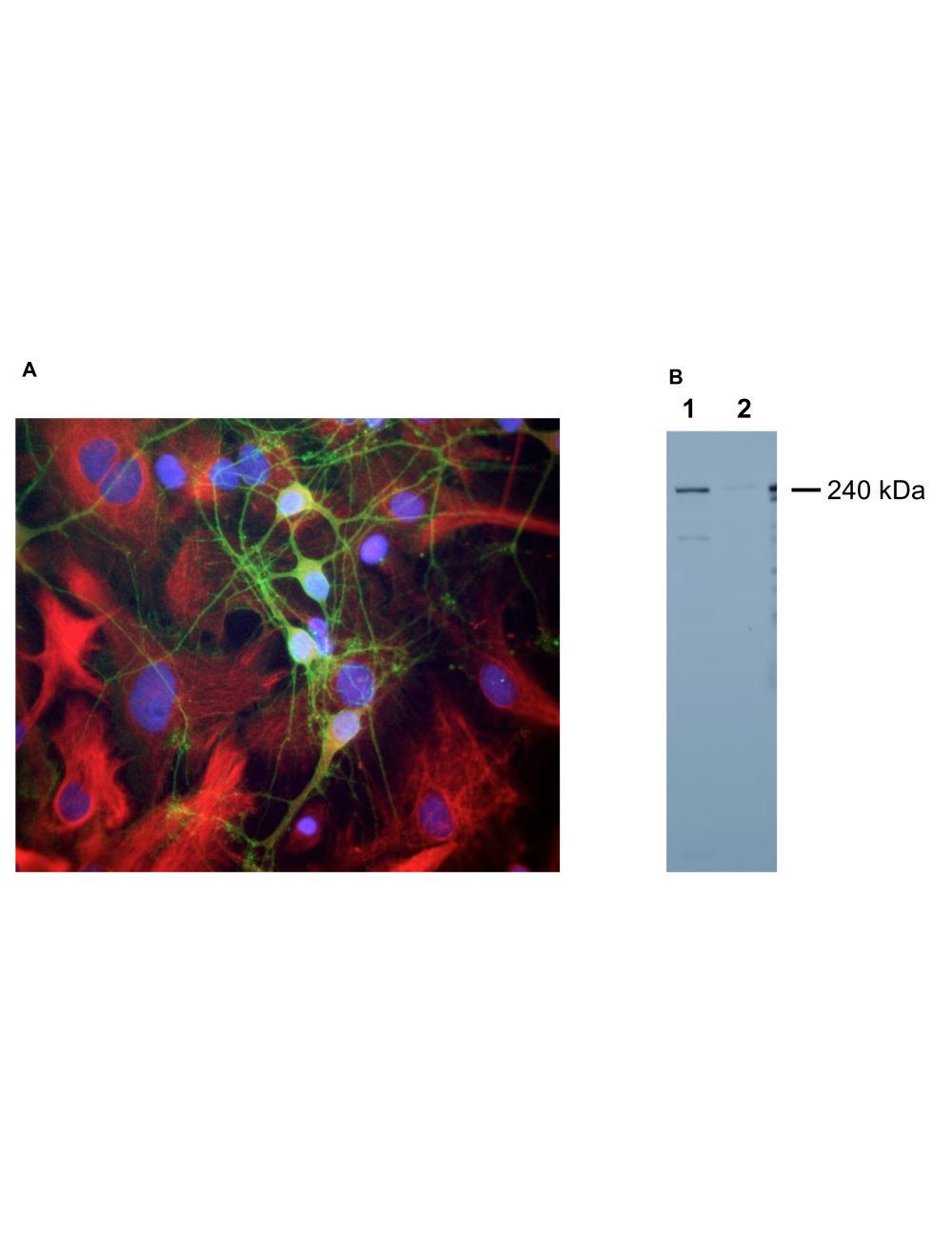

Spectrin, alpha II, Rabbit Polyclonal Antibody
As low as
US$317.00
Only %1 left
Catalog Number
R-1694
- Product Name Spectrin, alpha II, Rabbit Polyclonal Antibody
- Product Description Rabbit anti-Spectrin, alpha II Polyclonal Antibody (Unconjugated), suitable for WB, ICC.
- Application(s) ICC, WB
- Antibody Host Rabbit
- Antibody Type Polyclonal
- Specificity The antibody reacts with a 240 kDa band by Western blot on mouse sciatic nerve extract. Minor bands below may be seen and this likely represents in vivo proteolytic fragments of alpha-II spectrin. It has also been used successfully for immunocytochemistry.
- Species Reactivity Human, Mouse
- Immunogen Description The antibody was raised against a mix of five recombinant constructs containing the entire C-terminal region of human alpha-II spectrin (amino acids 676-2,400).
- Conjugate Unconjugated
- Purity Description Whole serum
- Regulatory Status For research use only.
Product Info
- Product Description Rabbit anti-Spectrin, alpha II Polyclonal Antibody (Unconjugated), suitable for WB, ICC.
- Application(s) ICC, WB
- Application Details Western Blotting (WB) and Immunocytochemistry (IC). Suggested dilution for WB is 1:5,000-10,000 and 1:500-1,000 for IC. Biosensis recommends optimal dilutions/concentrations should be determined by the end user.
- Target Spectrin, alpha II
- Specificity The antibody reacts with a 240 kDa band by Western blot on mouse sciatic nerve extract. Minor bands below may be seen and this likely represents in vivo proteolytic fragments of alpha-II spectrin. It has also been used successfully for immunocytochemistry.
- Target Host Species Human
- Species Reactivity Human, Mouse
- Antibody Host Rabbit
- Antibody Type Polyclonal
- Antibody Isotype IgG
- Conjugate Unconjugated
- Immunogen Description The antibody was raised against a mix of five recombinant constructs containing the entire C-terminal region of human alpha-II spectrin (amino acids 676-2,400).
- Purity Description Whole serum
- Format Lyophilized with sodium azide.
- Reconstitution Instructions Spin vial briefly before opening. Reconstitute with 100 µL sterile-filtered, ultrapure water. Centrifuge to remove any insoluble material.
- Storage Instructions Store lyophilized, unopened vial at 2-8°C or lower. After reconstitution, prepare aliquots and store at -20°C to -80°C for a higher stability. Avoid freeze-thaw cycles.
- Batch Number Please see item label.
- Expiration Date 12 months after date of receipt (unopened vial).
- Uniprot Number Q13813
- Uniprot Number/Name Q13813 (SPTN1_HUMAN)
- Scientific Background The spectrin family of proteins were originally discovered as major components of the submembraneous cytoskeleton of osmotically lysed red blood cells (1). The lysed blood cells could be seen as clear red blood cell shaped objects in the light microscope and were referred to as red cell "ghosts". The major proteins of these ghosts proved to be actin, ankyrin, band 4.1 and several other proteins, including two major bands running at about 240 kDa and 260 kDa on SDS-PAGE gels. This pair of bands was named "spectrin" since they were discovered in these red blood cell ghosts (1). Later work showed that similar high molecular bands were seen in membrane preparations from other eukaryotic cell types. Work by Levine and Willard described a pair of about ~240-260 kDa molecular weight bands which were transported at the slowest rate along mammalian axons (2). They named these proteins "fodrin" as antibody studies showed that they were localized in the sheath under the axonal membrane, but not in the core of the axon (2; fodros is Greek for sheath). Subsequently fodrin was found to be a member of the spectrin family of proteins, and the spectrin nomenclature is now normally used (3). Spectrins form tetramers of two alpha and two beta subunits, with the alpha corresponding to the lower molecular weight ~240 kDa band and the beta corresponding to the ~260 kDa or in some case much larger band. Most spectrin tetramers are about 0.2microns or 200nm long, and each alpha and beta subunit has a cell type specific expression pattern. The basic structure of each spectrin subunit is the spectrin repeat, which is a sequence of about 110 amino acids which defines a compact domain contain three closely packed alpha-helices. Each spectrin subunit contains multiple copies of this repeat, with 20 in each of the alpha subunits. The beta I-IV subunits each contain 17 spectrin repeats, while the beta V subunit, also known as beta-heavy spectrin, contains 30 of these repeats. The various subunits also contain several other kinds of functional domain, allowing the spectrin tetramer to interact with a variety of protein, ionic and lipid targets. The alpha-subunits each contain one calmodulin like calcium binding region and one Src-homology 3 (SH3) domain, an abundant domain involved in specific protein-protein interactions. The beta subunits all have a N-terminal actin binding domain and may also have one SH3 domain and one pleckstrin homology domain, a multifunctional type of binding domain which in beta I spectrin at least binds the membrane lipid PIP2 (5). Spectrins are believed to have a function in giving mechanical strength to the plasma membrane since the tetramers associate with each other to form a dense submembraneous geodesic meshwork (3). They also bind a variety of other membrane proteins and membrane lipids, and the proteins they bind to are therefore themselves localized in the membrane. Diseases may be associated with defects in one or other of the spectrin subunits (6). For example, some forms of hereditary spherocytosis, the presence of spherical red blood cells which are prone to lysis, can be traced to mutations in some of the spectrin subunits (7). The alpha-II subunit is widely expressed in tissues but, in the nervous system, is found predominantly in neurons. The antibody can therefore be used to identify neurons and fragments derived from neuronal membranes in cells in tissue culture and in sectioned material.
- Shipping Temperature 25°C (ambient)
- UNSPSC CODE 41116161
- Regulatory Status For research use only.
Specifications

-
General References
Marchesi VT & Steers E Jr. Selective solubilization of a protein component of the red cell membrane. Science 159:203-4 (1968).
Levine J & Willard M. Fodrin: axonally transported polypeptides associated with the internal periphery of many cells. J Cell Biol. 90:631-42 (1981).
Bennett V & Baines AJ. Spectrin and ankyrin-based pathways: metazoan inventions for integrating cells into tissues. Physiol Rev. 81:1353-92 (2001).
Djinovic-Carugo K, Gautel M, Ylaenne J & Young P. The spectrin repeat: a structural platform for cytoskeletal protein assemblies. FEBS Lett. 513:119-23 (2002).
Wang, DS and Shaw G. The association of the C-terminal region of beta I sigma II spectrin to brain membranes is mediated by a PH domain, does not require membrane proteins, and coincides with a inositol-1,4,5 triphosphate binding site. BBRC 217:608-15 (1995).
Bennett V & Healy J. Organizing the fluid membrane bilayer: diseases linked to spectrin and ankyrin. Trends Mol Med 14:28-36 (2008).Eber S & Lux SE. Hereditary spherocytosis--defects in proteins that connect the membrane skeleton to the lipid bilayer. Semin Hematol 41:118-41 (2004).