SpecificityClone LNC 1 recognizes an epitope on the outside of the regulatory N-terminus. The clone detects a protein of approximately 59-61 kDa by Western blot and reduced SDS-PAGE. The clone does not react with dopamine-beta-hydroxylase, phenylalanine hydroxylase, trytophan hydroxylase, dehydropteridine reductase, sepiapterin reductase or phenethanolamine-N-methyl transferase (PNMT) by western blots. Chicken, Frog, Horse, Human, Monkey, Mouse, Vole, Sheep, Zebrafish other species not yet tested
Species ReactivityChicken, Frog, Horse, Human, Mouse, Primate, Rat, Sheep, Vole, Zebra Fish
Immunogen DescriptionTyrosine Hydroxylase purified from PC12 cells
ConjugateUnconjugated
Purity DescriptionUnpurified ascites fluid, diluted with PBS containing 3% BSA, no preservatives.
Application DetailsWestern Blotting (WB), Immunohistochemistry (IHC), Immunohistochemistry/paraffin embedded IH(P), Immunoprecipitation (IP), Immunofluorescence (IF), Flow cytometry (FC).WB: 1:1000 -1:2000, SDS reduced samples. Detects a 59-61kDa protein. Rat Brain lysates is a suitable control. IHC/IH(P): Reacts in formalin fixed paraffin embedded tissues with HIER antigen recovery. Typical dilution is 1:100-1:200 depending upon incubation time and detection method used. IF: 1:200-1:1000, 4% PFA fixed tissues/cells permeabilized with 0.1-0.4% triton X-100; also works in fresh frozen and acetone fixed tissues/cells.IP: 1:100, immobilized on protein A beads, Fleming-Jones et al (1995) J. Protein Chemistry 14(5):275-282.FC: Fixed, permeabilized dopaminergic nerve terminals from rat striatum, {Wolf, ME, Kapatos, G (1989) The Journal of Neuroscience, January 1989, 9(l): 108-114; Wolf ME, Zigmond, MJ, Kapatos, G (1989) J. Neurochemistry 53(3):879-885}.
TargetTyrosine hydroxylase (TH)
SpecificityClone LNC 1 recognizes an epitope on the outside of the regulatory N-terminus. The clone detects a protein of approximately 59-61 kDa by Western blot and reduced SDS-PAGE. The clone does not react with dopamine-beta-hydroxylase, phenylalanine hydroxylase, trytophan hydroxylase, dehydropteridine reductase, sepiapterin reductase or phenethanolamine-N-methyl transferase (PNMT) by western blots. Chicken, Frog, Horse, Human, Monkey, Mouse, Vole, Sheep, Zebrafish other species not yet tested
Target Host SpeciesRat
Species ReactivityChicken, Frog, Horse, Human, Mouse, Primate, Rat, Sheep, Vole, Zebra Fish
Antibody HostMouse
Antibody TypeMonoclonal
Antibody IsotypeIgG1, kappa
Clone NameLNC, LNC1, LNC-1
ConjugateUnconjugated
Immunogen DescriptionTyrosine Hydroxylase purified from PC12 cells
Purity DescriptionUnpurified ascites fluid, diluted with PBS containing 3% BSA, no preservatives.
FormatLyophilized, dry powder.
Reconstitution InstructionsSpin vial briefly before opening. Reconstitute in 100 µL sterile-filtered, ultrapure water. Centrifuge to remove any insoluble material.
Storage InstructionsAfter reconstitution keep aliquots at -20 ° to -70°C for a higher stability. At 2-8°C keep up to one week, insulated, protected from light; use sterile methods and pipettes. Highly purified glycerol (1:1) may be added for an additional stability. Avoid repetitive freeze/thaw cycles. Keep tightly closed when not in use and protected from light
Batch NumberPlease see item label.
Expiration Date12 months after date of receipt (unopened vial).
Scientific BackgroundTyrosine hydroxylase is an excellent marker for dopaminergic and noradrenergic neurons. Tyrosine hydroxylase (a.k.a. tyrosine 3-monooxygenase) is the enzyme responsible for catalyzing the conversion of the amino acid L-tyrosine to L-3,4-dihydroxyphenylalanine (L-DOPA). L-DOPA is a precursor for dopamine, which, in turn, is a precursor for the important neurotransmitters norepinephrine (noradrenaline) and epinephrine (adrenaline). Tyrosine hydroxylase catalyzes the rate limiting step in this synthesis of catecholamines. In humans, tyrosine hydroxylase is encoded by the TH gene, and the enzyme is present in the central nervous system (CNS), peripheral symphatic neurons and the adrenal medulla. The enzymatic activity of TH requires ferrous ions as cofactors and is believed to be regulated by phosphorylation. At least four isoforms of human TH have been identified which result from alternative splicing. Tyrosine hydroxylase, phenylalanine hydroxylase and tryptophan hydroxylase together make up the family of aromatic amino acid hydroxylases (AAAHs).
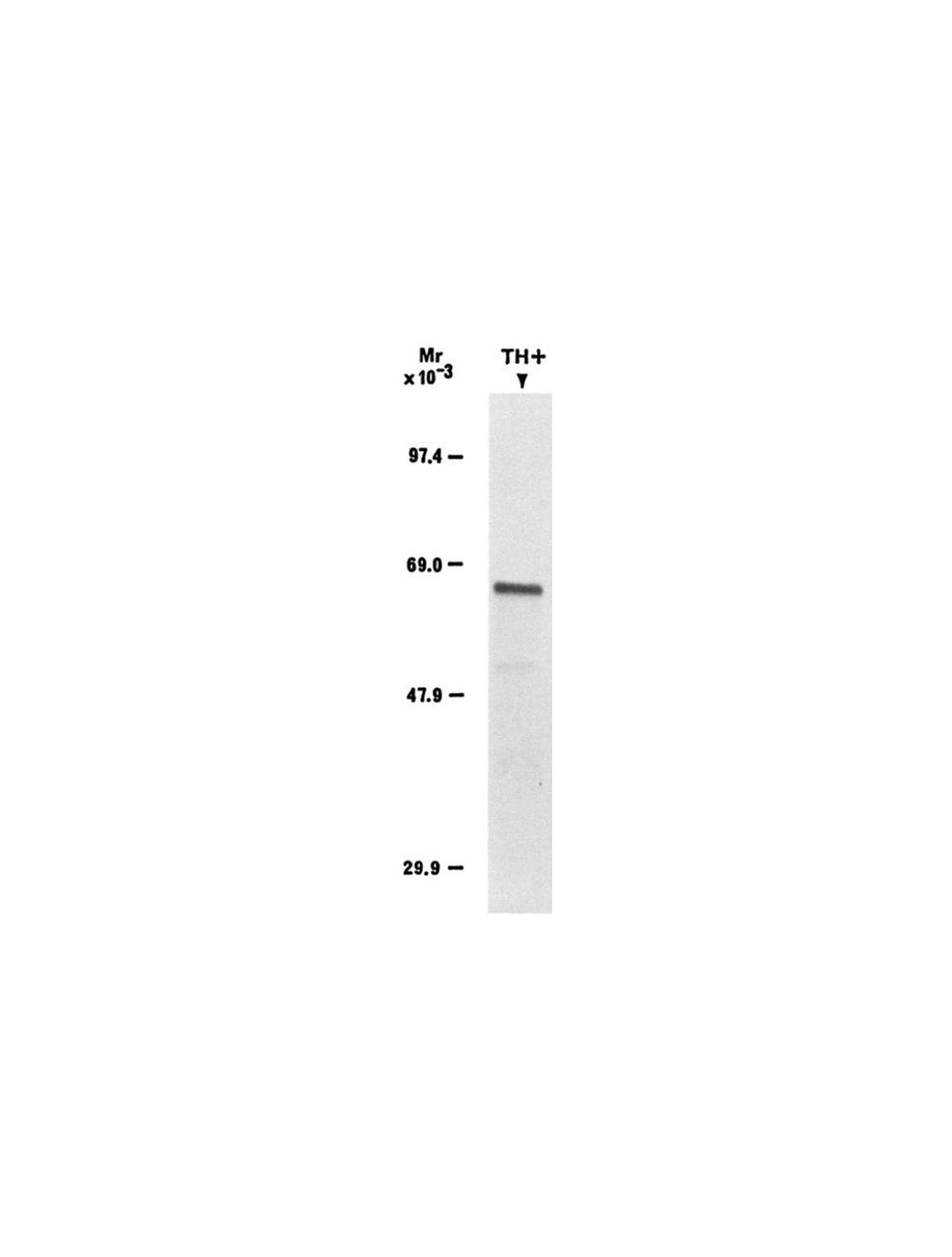
Approximately 0.5 µg of protein from purified synaptosomes analyzed by SDS-PAGE, electroblotted, and immunoprobed with LNC 1 Wolf, ME, Kapatos, G (1989) The Journal of Neuroscience, January 1989, 9(l): 108-114.
Staining of human substantia nigra with mouse monoclonal antibody to tyrosine hydroxylase, M-1616-100
Immunohistochemical detection of dopaminergic neurons in a formalin-fixed floated cryostat section from the human hypothalamus. The tyrosine hydroxylase primary antibody (M-1616-100; 1:50,000), was reacted with biotinylated secondary antibodies, followed by ABC Elite reagent (Vector). The peroxidase signal was visualized with nickel-diaminobenzidine. Photo courtesy of Dr. Erik Hrabovszky, Hungarian Academy of Sciences, Budapest, Hungary.
Specific ReferencesKapatos G., Kemski V., and Geddes T. (1989) Dopamine neurons in monolayer culture as a model system for the study of tyrosine hydroxylase, in Pteridines and Biogenic Amines in Neuropsychiatry, Pediatrics and Immunology (Levine R. A., KuhnD. M., Milstien S., and Curtius H-C., eds), pp. 243-258. Lakeshore Publishers, Grosse Pointe, Michigan.
 1800 605-5127
 +61 (0)8 8352 7711