Alternative NamesTyrosine hydroxylase; Tyrosine 3-monooxygenase; L-tyrosine hydroxylase; Tyrosine 3-hydroxylase;
Application(s)IHC-Frozen, WB
Antibody HostRabbit
Antibody TypePolyclonal
SpecificityIHC on brain shows a pattern of staining specific for TH containing neurons. This antibody is known to react with rat. Cross reactivity with other species has not yet been tested.
Species ReactivityHuman, Rat
Immunogen DescriptionA synthetic peptide (SPRFIGRRQSLIEDARK) as part of human Tyrosine Hydroxylase (aa: 32-47) conjugated to KLH
Application DetailsImmunohistochemistry (IHC): 0.5-1 ug/mL. This is a superb antibody for detection of tyrosine hydroxylase containing neurons exhibiting an intense labelling with a negligible background. This antiserum has proven extremely useful for staining of catecholaminergic neurons. It stains nicely and intensely dendritic processes and fine nerve terminals. Western Blotting (WB): 0.5-2 ug/mL. This antibody demonstrates clear immunoreactivity for TH at 60 kDa in rat PC12 cell lysate and mouse brain homogenate. Biosensis recommends optimal dilutions/concentrations should be determined by the end user.
TargetTyrosine Hydroxylase (TH)
SpecificityIHC on brain shows a pattern of staining specific for TH containing neurons. This antibody is known to react with rat. Cross reactivity with other species has not yet been tested.
Target Host SpeciesHuman
Species ReactivityHuman, Rat
Antibody HostRabbit
Antibody TypePolyclonal
Antibody IsotypeIgG
ConjugateUnconjugated
Immunogen DescriptionA synthetic peptide (SPRFIGRRQSLIEDARK) as part of human Tyrosine Hydroxylase (aa: 32-47) conjugated to KLH
Purity DescriptionAffinity purified
FormatLyophilized
Reconstitution InstructionsSpin vial briefly before opening. Reconstitute in 50 µL sterile-filtered, ultrapure water. Centrifuge to remove any insoluble material.
Storage InstructionsAfter reconstitution keep aliquots at -20°C for a higher stability, and at 2-8°C with an appropriate antibacterial agent. Glycerol (1:1) may be added for an additional stability. Avoid repetitive freeze/thaw cycles.
Batch NumberPlease see item label.
Expiration Date12 months after date of receipt (unopened vial).
Alternative NamesTyrosine hydroxylase; Tyrosine 3-monooxygenase; L-tyrosine hydroxylase; Tyrosine 3-hydroxylase;
Scientific BackgroundTyrosine hydroxylase (TH) is the rate-limiting enzyme in the synthesis of the catecholamines dopamine, epinephrine and norepinephrine. Therefore the regulation of the TH enzyme represents the central means for controlling the synthesis of these important catecholamines. FUNCTION: Plays an important role in the physiology of adrenergic neurons. CATALYTIC ACTIVITY: L-tyrosine + tetrahydrobiopterin + O2 = 3,4-dihydroxy-L-phenylalanine + 4a-hydroxytetrahydrobiopterin. COFACTOR: Fe(2+) ion. ENZYME REGULATION: Phosphorylation leads to an increase in the catalytic activity. PATHWAY: Catecholamine biosynthesis; first step. SUBUNIT: Homotetramer. PTM: In vitro, phosphorylation of Ser-19 increases the rate of Ser-40 phosphorylation, which results in enzyme opening and activation. SIMILARITY: Belongs to the biopterin-dependent aromatic amino acid hydroxylase family. The presence of different DNA sequences at the TH locus confers susceptibility to various disorders of the brain including manic-depression and schizophrenia. Parkinson's disease is also considered a TH deficiency as low dopamine levels are a consistent neurochemical abnormality.
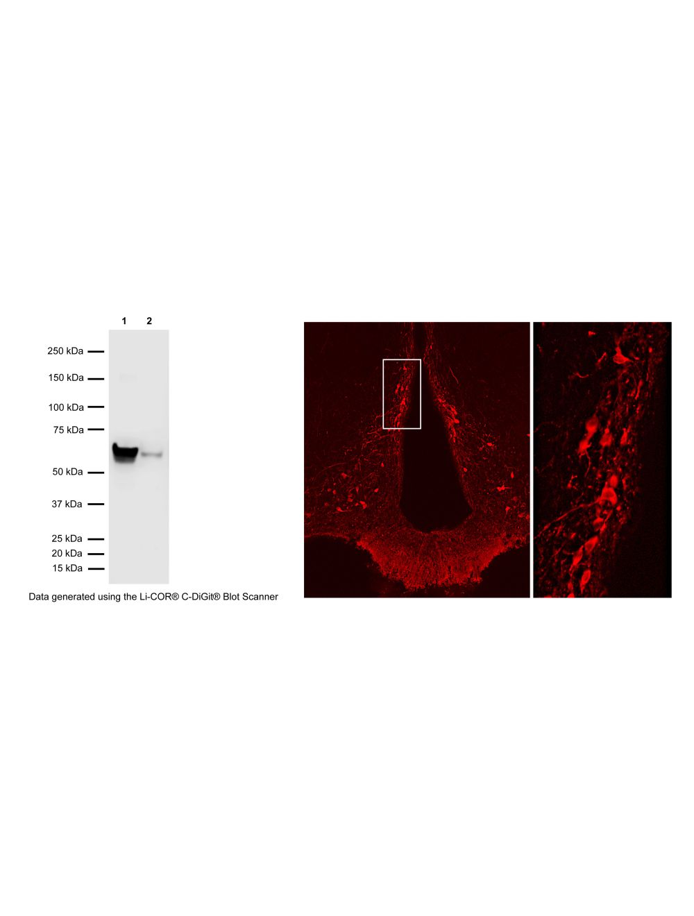
Left: Western Blot analysis of tyrosine hydroxylase (TH) expression in PC12 cell lysate (Lane 1: 6 ug) and mouse brain (Lane 2: 40 ug). The anti-TH rabbit antibody (1 µg/mL) detects one specific band at ~60 kDa corresponding to the expected molecular weight of TH in both samples. Method: SDS Page: denaturing and reducing, 4-12% Bis-Tris gel; Transfer: Tris-Glycine (Towbin's buffer) with 20% methanol; Membrane: PVDF (0.45 um); Blocking: 5% skim milk in TBST, 1 hr at RT; Primary antibody: R-148-50 (1 µg/mL), overnight at 2-8°C; Secondary antibody: donkey anti-rabbit (1:25,000), 1 hr at RT; Detection: Chemiluminescence. Right: Detection of TH-immunoreactivity in tuberoinfundibular dopaminergic neurons in rat hypothalamic arcuate nucleus by Immunohistochemistry. Primary antibody: rabbit anti-TH cat# R-148-50 (1:2,000); Secondary antibody: Cy3-conjugated anti-rabbit. Photo courtesy of Dr. Erik Hrabovszky, Hungarian Academy of Sciences, Budapest, Hungary.
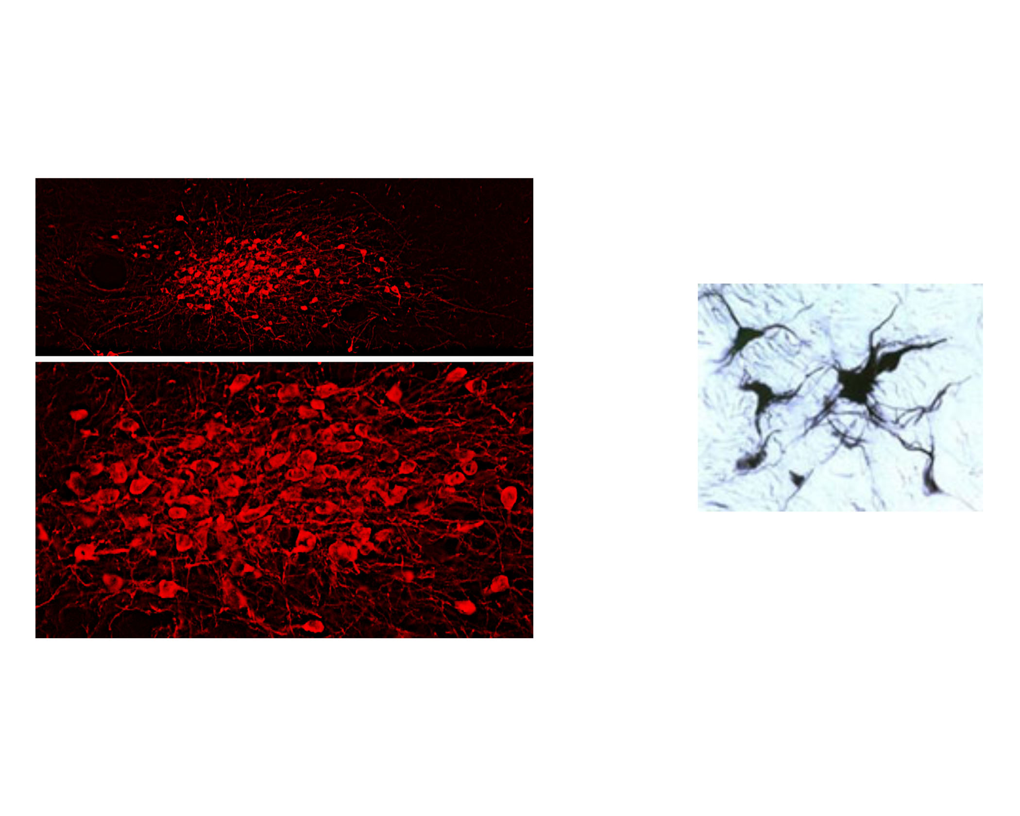
Left: Visualization of dopaminergic neurons in formalin-fixed floated cryostat section from the rat zona incerta by Immunohistochemistry. TH-immunoreactivity was detected with rabbit anti-TH antibody R-148-50 (1:2,000), followed by Cy3-conjugated anti-rabbit antibody. Photo courtesy of Dr. Erik Hrabovszky, Hungarian Academy of Sciences, Budapest, Hungary. Right: Staining of TH-IR catacholaminergic neurons in the rat brainstem using the DAB method.
General ReferencesMallett, J. Trends in Pharmacological Science. 17(4): 129-135, 1996. Haavik, J. et al. Mol. Neurobiology 16(3) :285-309, 199 Lewis DA, et al, Neuroscience 54: 477-, 1993 Kumer S.C. et al. Journal of Neurochemistry, 67(2) :443-462, 199 Haycock, J. Anal. Biochemistry 181: 259-266, 198 Haycock, J. Anal. Biochemistry 208: 397-399, 199 Renfroe, J.B., et al. Brain Res. Bull. 13: 109 - 126, 198 Xu, Z et a.l Neurosci. 82(3): 727, 1998
 1800 605-5127
 +61 (0)8 8352 7711