Product NameTyrosine Kinase Receptor A (TrkA), Mouse Monoclonal Antibody
Product DescriptiongoogleMouse anti-Tyrosine Kinase Receptor A (TrkA) Monoclonal Antibody (Unconjugated), suitable for ICC, FC.
Alternative NamesTropomyosin-related kinase receptor; High affinity nerve growth factor receptor; Neurotrophic tyrosine kinase receptor type 1; TRK1 transforming tyrosine kinase protein; p140-TrkA; Trk-A; NTRK1; TRK
Application(s)FC, ICC
Antibody HostMouse
Antibody TypeMonoclonal
SpecificityTrkA. Does not cross-react with TrkB or TrkC. Reacts with human TrkA. While not tested yet, M-1723-100 is not expected to react with TrkA from other species due to amino acid sequence dissimilarity.
Species ReactivityHuman
Immunogen DescriptionA synthetic peptide from the extracellular domain of human TrkA (C-SATVMKSGGLPS, aa: 235-246) has been used as the immunogen.
ConjugateUnconjugated
Purity DescriptionProtein G purified mouse immunoglobulin
Product DescriptionMouse anti-Tyrosine Kinase Receptor A (TrkA) Monoclonal Antibody (Unconjugated), suitable for ICC, FC.
Application(s)FC, ICC
Application DetailsFlow cytometry: 20 µg/mL.
Immunocytochemistry: 1-5 µg/mL.
Flow cytometry data and immunofluorescence staining of TrkA (extracellular domain) receptor in SHSY-5Y cells has shown that detection of TrkA expression depends on its cellular localization (membrane vs. internal stores). Thus, permeabilization of cells may be required, despite that clone BS470 was raised against the extracellular domain of TrkA.
Other applications not yet tested. Biosensis recommends optimal dilutions/concentrations should be determined by the end user.
TargetTyrosine Kinase Receptor A (TrkA)
SpecificityTrkA. Does not cross-react with TrkB or TrkC. Reacts with human TrkA. While not tested yet, M-1723-100 is not expected to react with TrkA from other species due to amino acid sequence dissimilarity.
Target Host SpeciesHuman
Species ReactivityHuman
Antibody HostMouse
Antibody TypeMonoclonal
Antibody IsotypeIgG3, kappa
Clone NameBS470
ConjugateUnconjugated
Immunogen DescriptionA synthetic peptide from the extracellular domain of human TrkA (C-SATVMKSGGLPS, aa: 235-246) has been used as the immunogen.
SequenceSATVMKSGGLPS
Purity DescriptionProtein G purified mouse immunoglobulin
FormatLyophilized from PBS, pH 7.4, containing 3% trehalose without preservatives.
Reconstitution InstructionsSpin vial briefly before opening. Reconstitute in 100 µL sterile-filtered, ultrapure water. Centrifuge to remove any insoluble material. Final buffer contains no preservatives.
Storage InstructionsAfter reconstitution divide into aliquots and store at -20°C for a higher stability. Antibody contains no preservatives. Store at 2-8°C with an appropriate antibacterial agent. Use sterile methods. Highest purity Glycerol (1:1) may be added for an additional stability when stored at refrigerated or freezing temperatures. Avoid repetitive freeze/thaw cycles.
Batch NumberPlease see item label.
Expiration Date12 months after date of receipt (unopened vial).
Alternative NamesTropomyosin-related kinase receptor; High affinity nerve growth factor receptor; Neurotrophic tyrosine kinase receptor type 1; TRK1 transforming tyrosine kinase protein; p140-TrkA; Trk-A; NTRK1; TRK
Scientific BackgroundTrkA is a member of the neurotrophic tyrosine kinase receptor family. It is a membrane-bound receptor that upon neurotrophin binding, phosphorylates itself and members of the MAPK pathway. TrkA is required for high-affinity binding to nerve growth factor (NGF), neurotrophin-3 and neurotrophin-4/5 but not brain-derived neurotrophic factor (BDNF). TrkA leads to cell differentiations and may play a role in specifying sensory neuron subtypes. It has a crucial role in the development and function of the nociceptive reception system as well as establishment of thermal regulation via sweating. SUBUNIT: Exists in a dynamic equilibrium between monomeric (low affinity) and dimeric (high affinity) structures. SUBCELLULAR LOCATION: Cell membrane; single-pass type I membrane protein. Endocytosed to the endosomes upon treatment of cells with NGF. ALTERNATIVE PRODUCTS: 2 named isoforms produced by alternative splicing. Both isoforms have similar biological properties. TISSUE SPECIFICITY: Isoform TrkA-II is primarily expressed in neuronal cells. Isoform TrkA-I is found in non-neuronal tissues. Mutations in TrkA have been associated with congenital insensitivity to pain, anhidrosis, self-mutalating behaviour, mental retardation and cancer (Reference: www.uniprot.com).
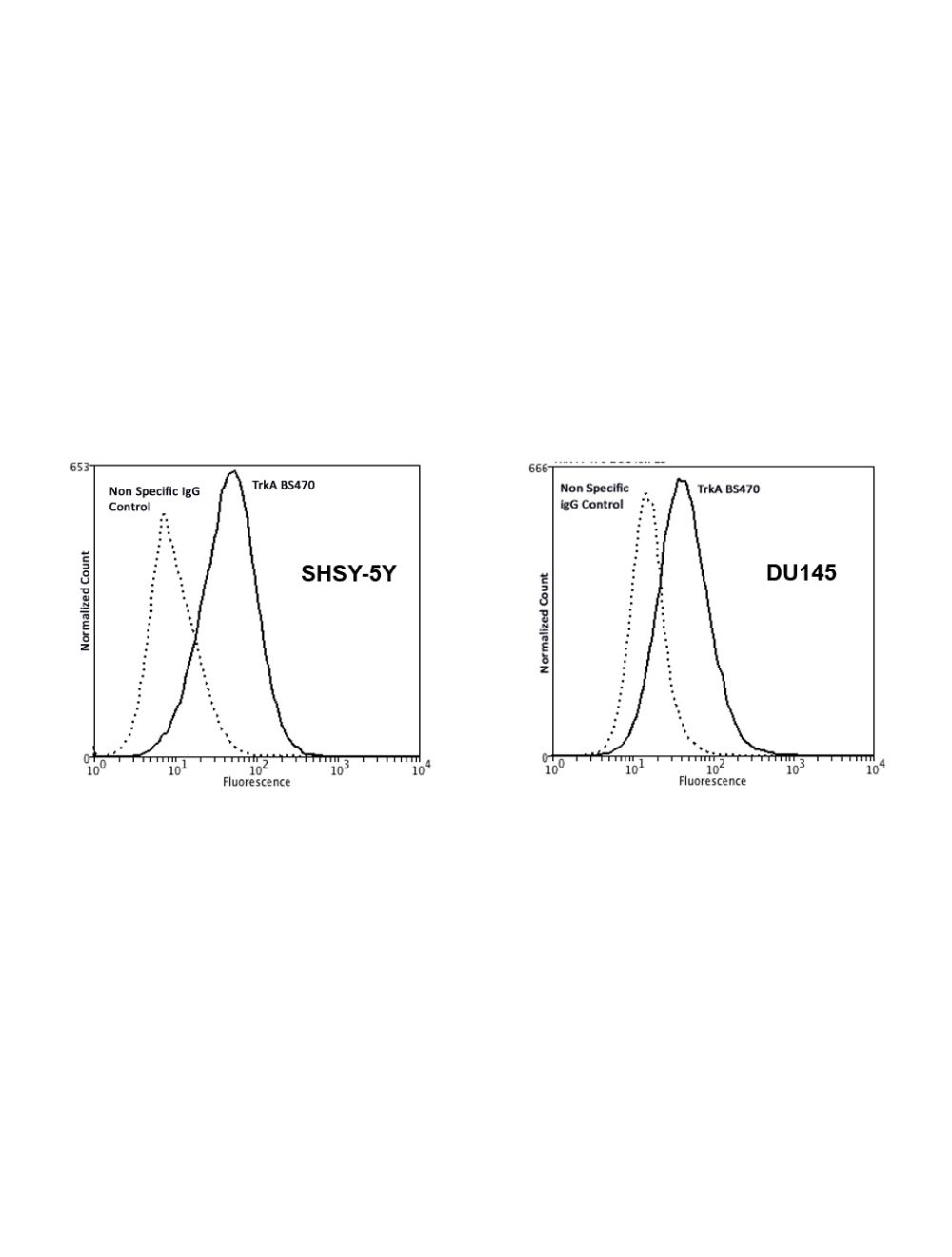
TrkA expression on non-permeabilized human SHSY-5Y neuroblastoma and DU145 prostate cancer cell lines analysed by Flow Cytometry. Blocking: 200 µg/mL normal sheep IgG (30 minutes) on ice. Primary antibody: Mouse Monoclonal antibody to TrkA, extracellular domain (cat # M-1723-100, 2 μg per ~106 cells) for 30 minutes on ice. Secondary antibody: Goat anti-mouse PE (1:100 dilution, 20 min in dark on ice. Non-specific Control IgG, clone X63 (cat# M-1249-100) was used as negative control under same conditions (black dashed). Flow cytometry data and results were generated using Orflo Moxiflow™ instrument and protocols.
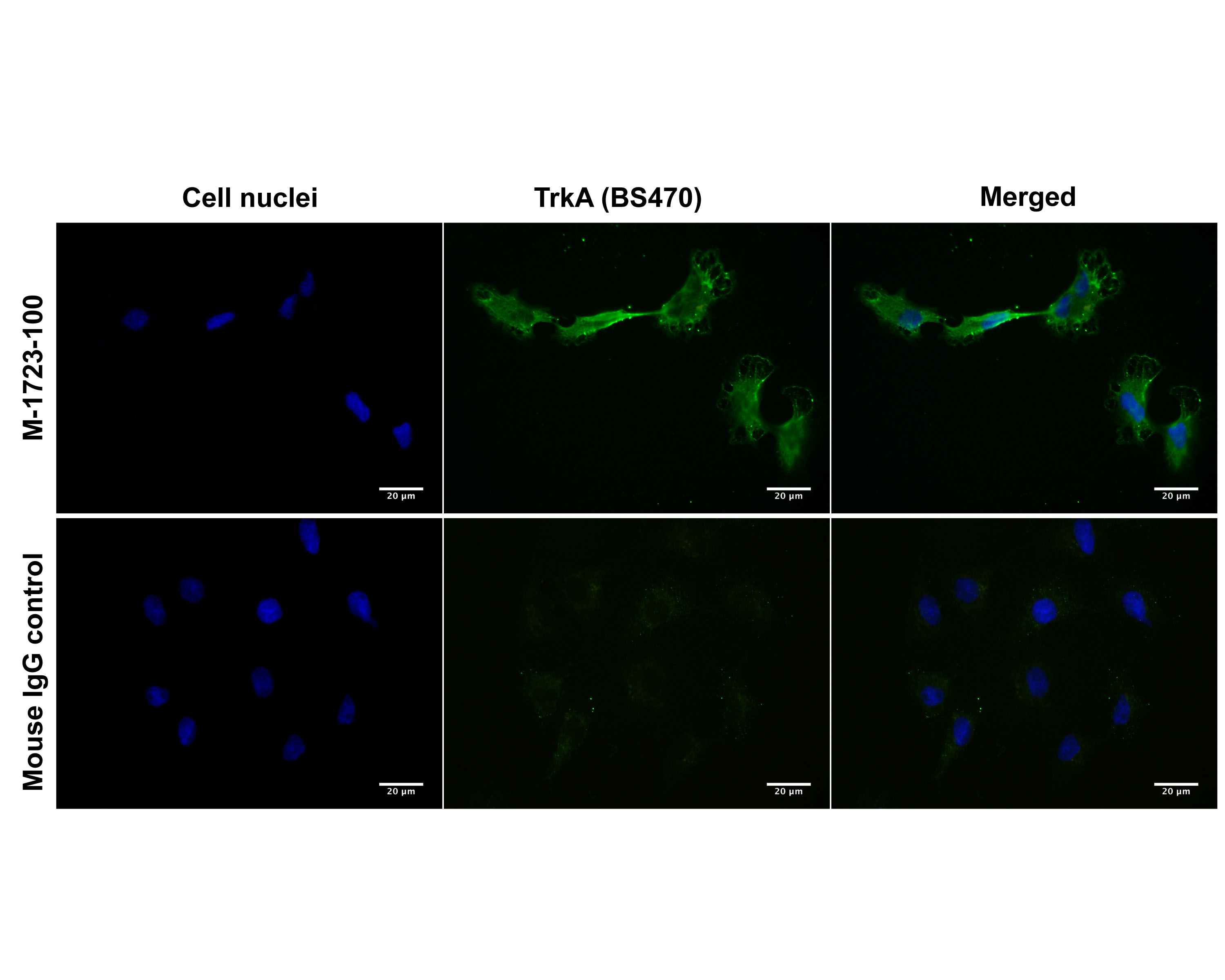
Immunofluorescence analysis of TrkA expression on human SHSY-5Y neuroblastoma cell membrane. Fixed (4% formaldehyde), non-permeabilized, and blocked (10% normal horse serum) SHSY-5Y cells were incubated with TrkA antibody M-1723-100 (extracellular domain, 2 µg/mL, green) for 1 hour. Primary antibody binding was visualized with a secondary donkey anti-mouse-CF488A antibody (4 µg/mL, 1 hour incubation). Cell nuclei were stained with Hoechst dye (blue). Magnification: 100x.
Immunofluorescence analysis of TrkA expression in human SHSY-5Y neuroblastoma cells, comparing TrkA antibodies M-1723-100 (BS470, extracellular domain) and M-1719-100 (BS292, intracellular domain). Cells were fixed (4% formaldehyde, 10 minutes), permeabilized (0.1% Triton X100, A and C) or non-permeabilized (A), and blocked (10% normal horse serum) for 30 minutes. SHSY-5Y cells were incubated for 1 hour with TrkA primary antibodies (2 µg/mL, green) M-1723-100 (extracellular domain, A1 and B1) and M-1719-100 (intracellular domain, C1). Primary antibody binding was visualized with a secondary donkey anti-mouse-CF488A antibody (4 µg/mL, 1 hour incubation). Cell nuclei were stained with Hoechst dye (blue). Control cells were treated exactly the same way, substituting the primary TrkA antibody with normal mouse IgG (A2, B2 and C2) and using the same camera exposure times to record images. Magnification: 100x. This batch of SHSY-5Y cells did not show TrkA expression on the cell membrane (A), while both antibodies were immunoreactive for intracellulary stored TrkA receptor (B and C).
Comparison of TrkA expression on non-permeabilized and permeabilized human SHSY-5Y neuroblastoma cells by Flow Cytometry. This batch of SHSY-5Y cells did not show TrkA expression on the membrane (left image), but immunoreactivity in permeabilized cells (right image), suggesting that TrkA is stored intracellularly. Conditions: Permeabilization with absolute methanol (10 minutes on ice), or no permeabilization (incubation of primary antibody on ice only). Blocking: 200 µg/mL normal sheep IgG (30 minutes) on ice. Primary antibody: Mouse Monoclonal antibody to TrkA, extracellular domain (cat# M-1723-100, 2 µg per ~106 cells) for 30 minutes on ice. Secondary antibody: Goat anti-mouse PE (1:100 dilution, 20 min in dark on ice. Non-specific Control IgG, clone X63 (cat# M-1249-100) was used as negative control under same conditions (black dashed). Flow cytometry data and results were generated using Orflo MoxiflowTM instrument and protocols.
 1800 605-5127
 +61 (0)8 8352 7711