Product NameTyrosine Kinase Receptor B (TrkB), Rabbit Polyclonal Antibody
Product DescriptiongoogleRabbit anti-Tyrosine Kinase Receptor B (TrkB) Polyclonal Antibody (Unconjugated), suitable for WB, FC.
Alternative NamesTrk-B,TrkB, GP145-TrkB, Neurotrophic tyrosine kinase receptor type 2, TrkB tyrosine kinase, Tropomyosin-related kinase B
Application(s)FC, WB
Antibody HostRabbit
Antibody TypePolyclonal
SpecificityTrkB; detects TrkB in both human, mouse and rat samples by western blot and has positive staining by Flow cytometry against human TrkB positive cell lines.
Application DetailsWestern Blotting (0.1 - 0.5 µg/mL): Tested on RIPA lysates from human and rodent brain tissue. Detects TrkB full-length (~140 kDa) and truncated TrkB (~100 kDa) in tissue homogenates. In cell lysates, only a ~50 kDa uncharacterized TrkB isoform is detected. Flow Cytometry (5-20 µg/mL): Tested on human cell lines.
Other applications have not been tested. Biosensis recommends optimal dilutions/concentrations should be determined by the end user.
TargetTyrosine Kinase Receptor B (TrkB)
SpecificityTrkB; detects TrkB in both human, mouse and rat samples by western blot and has positive staining by Flow cytometry against human TrkB positive cell lines.
Positive ControlRat or mouse cortex (WB). SHSY5Y cell line (FC).
Purity DescriptionAffinity purified
FormatLyophilized from a solution containing 5 mg BSA, 0.9 mg NaCl, 0.2 mg Na2HPO4, 0.05 mg Thimerosal, 0.05 mg NaN3.
Reconstitution InstructionsSpin vial briefly before opening. Reconstitute in 100 uL sterile-filtered, ultrapure water to obtain an antibody concentration of 1 mg/mL. Centrifuge to remove any insoluble material.
Storage InstructionsStore lyophilized antibody at 2-8°C. After reconstitution divide into aliquots and store at -20°C for long-term storage. Store at 2-8°C short-term (up to 4 weeks). Avoid repetitive freeze/thaw cycles.
Batch NumberPlease see item label.
Expiration Date12 months after date of receipt (unopened vial).
Alternative NamesTrk-B,TrkB, GP145-TrkB, Neurotrophic tyrosine kinase receptor type 2, TrkB tyrosine kinase, Tropomyosin-related kinase B
Scientific BackgroundThe protein named TrkB (also named Neurotrophic tyrosine kinase receptor type 2 (NTRK2), GP145-TrkB or Tropomyosin-related kinase B is a receptor tyrosine kinase involved in the development and the maturation of the central and the peripheral nervous systems and is important in the regulation of neuron survival, proliferation, migration, differentiation, and synapse formation and plasticity. TrkB may also play a role in neutrophin-dependent calcium signaling in glial cells and mediate communication between neurons and glia. TrkB is the primary receptor for BDNF (brain-derived neurotrophic factor. TrkB also binds NT4 and NT3 but less efficiently. Upon ligand-binding, the receptor undergoes homodimerization, autophosphorylation and activation. TrkB activation recruits, phosphorylates and/or activates several downstream effectors including SHC1, FRS2, SH2B1, SH2B2 and PLCG1 that each regulate distinct overlapping signaling cascades within cells. Through SHC1, FRS2, SH2B1, SH2B2, these activate the GRB2-Ras-MAPK cascade that regulates, for instance, neuronal differentiation including neurite outgrowth. These same effectors also control the Ras-PI3 kinase-AKT1 signaling cascade that mainly regulates growth and survival. TrkB, via activation of PLCG1 and the downstream protein kinase C-regulated pathways, also controls synaptic plasticity, and thus plays a role in learning and memory by regulating both short term synaptic function and long-term potentiation. PLCG1 also leads to NF-Kappa-B activation and the transcription of genes involved in cell survival. One such consequence is that PLCG1 activation via TrkB is able to suppress anoikis, the apoptosis resulting from loss of cell-matrix interactions. (Reference: www.uniprot.org)
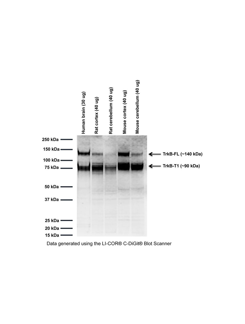
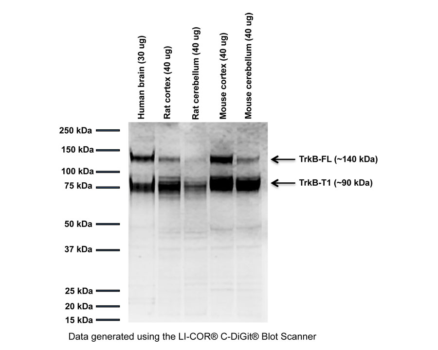
Western blot analysis of TrkB expression in various human and rodent tissue homogenates (RIPA extracts). Two prominent bands are detected at ~140 kDa and ~90 kDa, corresponding to TrkB full-length (FL) and truncated TrkB (T1) isoforms. SDS-PAGE: denatured and reduced; Transfer: Towbin's buffer (20% MeOH, no SDS); Membrane: PVDF (0.45 um); Blocking: 5% skim milk in TBST, 1 hour at RT; Primary antibody: overnight at 2-8°C (0.5 µg/mL); Secondary antibody: anti-rabbit-HRP (1/10,000), 1 hours at RT; Detection: Chemiluminiscence.
Analysis of membrane TrkB expression in three human cell lines by Flow Cytometry. Staining was perfomed under native conditions. Blocking: 10% normal horse serum; Primary antibody: R-1834-100 (red), 10 µg/mL, 30 minutes on ice; Secondary antibody: donkey anti-rabbit PE (1:100), 30 minutes in dark on ice. Negative control: normal rabbit IgG (black). Data and results were generated using Orflo MoxiflowTM instrument and protocols.
Western Blot and Immunoprecipitation analysis of TrkB expression in cell lysate and tissue homogenates. Rabbit anti-TrkB antibody detects two main TrkB isoforms at ~140 kDa (full-length) and ~100 kDa (truncated TrkB, TrkB-T1) in tissue homogenates (red arrows). In cell lysate, a TrkB isoform at ~50 kDa is observed (black arrow) which is immunoprecipitated with mouse anti-TrkB clone BS379 (M-1836-100). Additional uncharacterized bands are observed. Western Blotting Method: SDS-PAGE: denaturing and reducing, 4-12% Bis-Tris gel; Transfer: Towbin’s transfer buffer; Membrane: PVDF (0.45 µm); Blocking: 5% skim milk in TBS pH 7.6 (1 hour at RT); Primary antibody: rabbit anti-TrkB (0.5 µg/mL), overnight at 2-8°C; Secondary antibody: anti-rabbit-HRP (1/10,000), 1 hour at RT; Detection: ECL. Immunoprecipitation Method: 2 mg protein lysate in non-denaturing buffer (20 mM Tris, 137 mM NaCl, 1% Triton X-100, 2 mM EDTA, protease inhibitor cocktail) was pre-cleared with Protein G agarose (80 uL of 50% bead slurry) and TrkB precipitated from 500 ug protein lysate with 20 µg TrkB antibody clone BS379 (M-1836-100), or negative control IgG clone X63 (M-1249-100). Immunocomplexes were captured with Protein G agarose (10 uL of 50% bead slurry) and eluted with 25 µL of 4x sample buffer. Precipitated proteins were subjected to Western Blotting as outlined above.
Western Blot analysis of TrkB expression in RIPA cell lysates. Rabbit anti-TrkB antibody detects one TrkB isoforms at ~50 kDa which is immunoprecipitated with mouse antibody to TrkB (M-1836-100). In all cell lysates tested, R-1834-100 does not detect ~140 kDa TrkB (full-length) or ~100 kDa (truncated TrkB, TrkB-T1). Western Blotting Method: SDS-PAGE: denaturing and reducing, 10% Bis-Tris gel; Transfer: Towbin’s Transfer buffer; Membrane: PVDF (0.45 µm); Blocking: 5% skim milk in TBS pH 7.6 (1 hour at RT); Primary antibody: rabbit anti-TrkB (0.5 µg/mL), overnight at 2-8°C; Secondary antibody: anti-rabbit-HRP (1/10,000), 1 hour at RT; Detection: ECL.
Specific ReferencesFleury S et al. (2021) Tissue-Specificity of Antibodies Raised Against TrkB and p75NTR Receptors; Implications for Platelets as Models of Neurodegenerative Diseases. Front Immunol. 12:606861Application: WB, human platelets.
 1800 605-5127
 +61 (0)8 8352 7711