Alternative NamesPro-brain nerve growth factor; proNGF; NGF
Application(s)FC, ICC, WB
Antibody HostMouse
Antibody TypeMonoclonal
SpecificityHuman Species cross-reactivity not tested.
Species ReactivityHuman
Immunogen DescriptionA synthetic peptide (C-HTIPQAHWTKLQ, aa: 30-41) of human proNGF protein has been used as the immunogen. The sequence is located on the pro-domain of the proNGF full-length protein and is 80% homologous to mouse and rat proNGF.
Application DetailsFlow Cytometry (2 ug/ 10^6 cells). Immunocytochemistry (1-2 µg/mL), Western Blotting (1-2 µg/mL). Other applications not yet tested. Biosensis recommends optimal dilutions/concentrations should be determined by the end user.
TargetPro-nerve growth factor (proNGF)
SpecificityHuman Species cross-reactivity not tested.
Target Host SpeciesHuman
Species ReactivityHuman
Antibody HostMouse
Antibody TypeMonoclonal
Antibody IsotypeIgG2b, lambda
Clone NameBS312
ConjugateUnconjugated
Immunogen DescriptionA synthetic peptide (C-HTIPQAHWTKLQ, aa: 30-41) of human proNGF protein has been used as the immunogen. The sequence is located on the pro-domain of the proNGF full-length protein and is 80% homologous to mouse and rat proNGF.
Purity DescriptionProtein G purified mouse IgG.
FormatLyophilized from a solution containing PBS buffer pH 7.4 with 3% trehalose, without preservatives.
Reconstitution InstructionsSpin vial briefly before opening. Reconstitute in 100 µL sterile-filtered, ultrapure water. Centrifuge to remove any insoluble material. Final buffer contains no preservatives.
Storage InstructionsStore lyophilized antibody at 2-8ºC. After reconstitution divide into aliquots and store at -20°C for a higher stability. Antibody contains no preservatives. Store at 2-8°C with an appropriate antibacterial agent. Use sterile methods. Highest purity Glycerol (1:1) may be added for an additional stability when stored at refrigerated or freezing temperatures. Avoid repetitive freeze/thaw cycles.
Batch NumberPlease see item label.
Expiration Date12 months after date of receipt (unopened vial).
Alternative NamesPro-brain nerve growth factor; proNGF; NGF
Scientific BackgroundNerve growth factor (NGF) is synthesized as a precursor (proNGF) which may be released and have physiological functions to cause cell death. It binds neurotrophin receptor p75 and sortilin and may also be important for the development of nervous system. proNGF is synthesized in target tissues and glia, transported retrogradely and may be released.
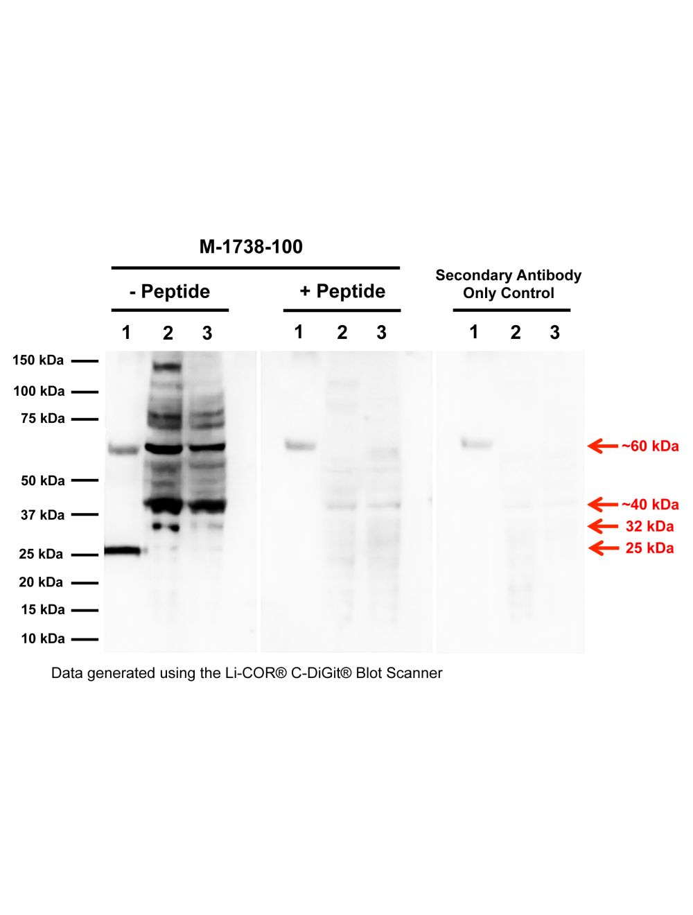
Western blot analysis of proNGF expression in DU145 and PC3 prostate cancer cell lysates. Monoclonal proNGF antibody M-1738-100 detects monomeric, non-glycosylated proNGF protein (20 ng, E.coli derived) at 25 kDa (Sample 1). PC3 (Sample 2, 100 µg) and DU145 (Sample 3, 100 µg) cell lysates show several proNGF isoforms (32 kDa, ~40 kDa, ~60 kDa, and higher molecular weight bands) as previously described, which most likely relates to glycosylated forms of full-length proNGF (Reinshagen et al., 2000; Lobos et al., 2005; Pedraza et al., 2005; Pundavela et al., 2014). Specificity of these bands is shown by pre-incubation with the immunizing peptide (5x mass of primary antibody), and a secondary antibody only control blot. Western Blotting Method: SDS-PAGE: denaturing and reducing, 4-20% Bis-Tris gel; Semi-Dry Transfer: Tris-Glycine (Towbin's) buffer with 20% methanol; Membrane: Nitrocellulose (0.45 µm); Blocking: 5% skim milk in TBST, 1 hour at RT; Primary antibody: 2 µg/mL, overnight at 4°C; Secondary antibody: anti-mouse-HRP (1/6000), 1 hour at RT; Detection: Chemiluminiscence.
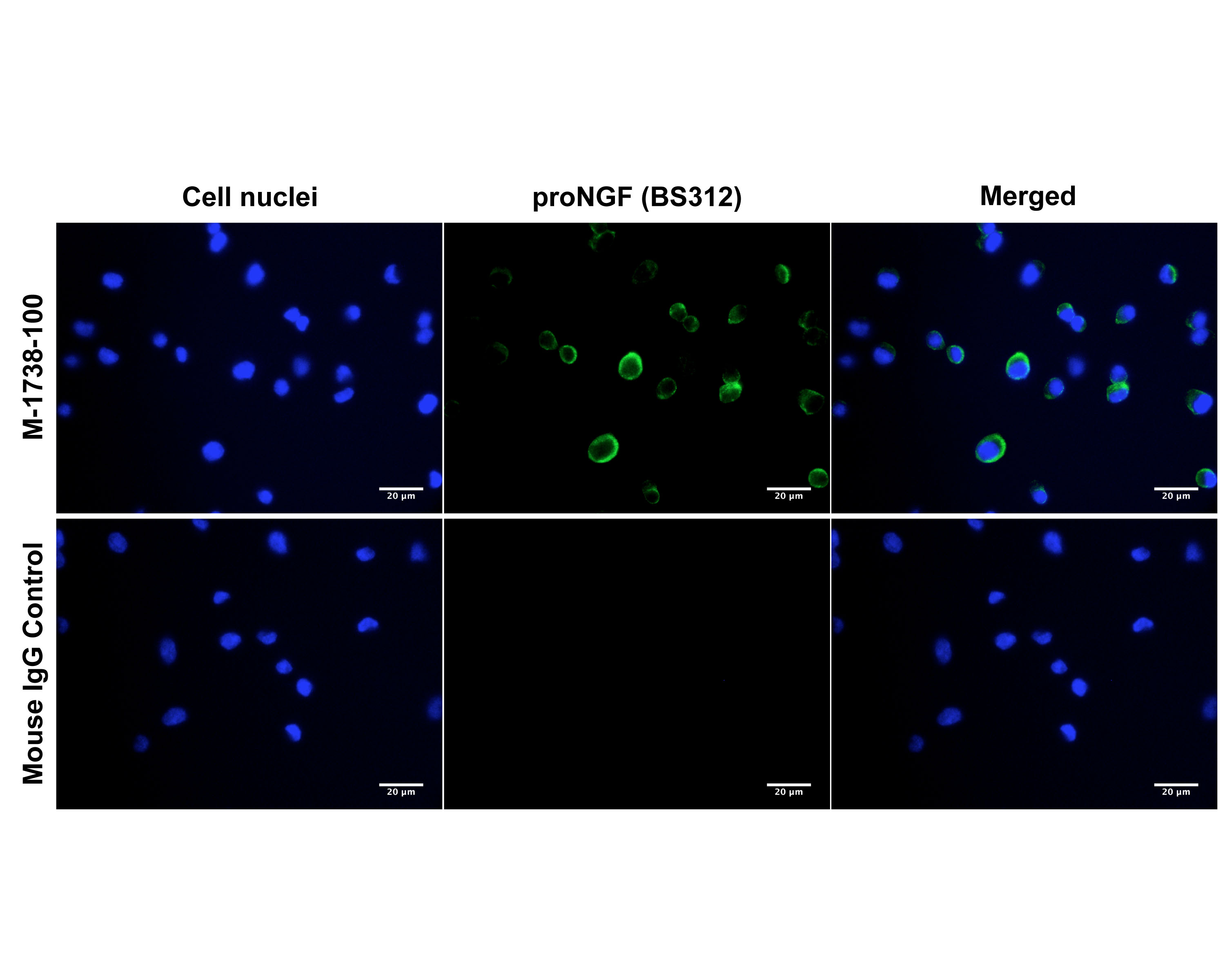
Immunofluorescence analysis of proNGF expression in human DU145 prostate cancer cells. Fixed (4% formaldehyde), permeabilized, and blocked (10% normal horse serum, 0.1% Triton X100) DU145 cells were incubated with proNGF antibody M-1738-100 (2 µg/mL, green) for 1 hour. Primary antibody binding was visualized with a secondary donkey anti-mouse-CF488A antibody (4 µg/mL, 1 hour incubation). Cell nuclei were stained with Hoechst dye (blue). Magnification: 100x.
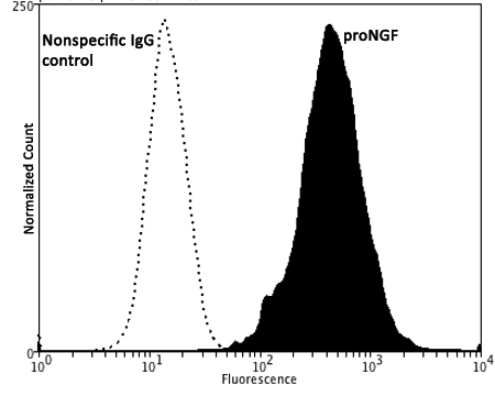
Fixing and Permeabilization of cells: Absolute methanol (10 minutes in ice) and 0.1% Tween-20 in PBS, Blocking: 200 µg/mL Normal Sheep IgG (20 minutes), Primary antibody: Mouse Monoclonal antibody to proNGF (cat # M-1738-100, 2 ug per ~10^6 cells) for 60 minutes at room temperature, Secondary antibody: Goat anti-mouse PE labeled secondary antibody (1:100 fold dilution) with incubation for 20 minutes in dark at room temperature. Non-specific Control IgG, clone X63 (cat # M-1249-100) was used as negative control under same conditions (black dashed). Flow cytometry data and results were generated using Orflo MoxiflowTM instrument and protocols. The data demonstrates specific staining of proNGF expressed in human prostate cancer DU145 cell line using cat# M-1738-100.
Western blot analysis of proNGF expression in human brain homogenates. Monoclonal proNGF antibody M-1738-100 (2 µg/mL) detects monomeric, non-glycosylated proNGF protein (30 ng, E.coli-derived) at 25 kDa (Sample 1). Brain homogenate 1 (Sample 2, 50 ug) and 2 (Sample 3, 30 ug) show several proNGF isoforms (~53 kDa, ~60 kDa, and ~73 kDa) as previously described, which most likely relates to glycosylated forms of full-length proNGF (Reinshagen et al., 2000; Lobos et al., 2005; Pedraza et al., 2005; Pundavela et al., 2014). Specificity of these bands is shown by pre-incubation with the immunizing peptide (5x mass of primary antibody). Western Blotting Method: SDS-PAGE: denaturing and reducing, 4-20% Bis-Tris gel; Semi-Dry Transfer: Tris-Glycine (Towbin's) buffer with 20% methanol; Membrane: Nitrocellulose (0.45 um); Blocking: 5% skim milk in TBST, 1 hour at RT; Primary antibody: overnight at 4°C; Secondary antibody: anti-mouse-HRP (1/6000), 1 hour at RT; Detection: Chemiluminiscence.
Specific ReferencesMarsland M et al. (2023). "ProNGF Expression and Targeting in Glioblastoma Multiforme." Int J Mol Sci. 24(2):1616 Application: WB, Invasion assay
 1800 605-5127
 +61 (0)8 8352 7711