SpecificityLess than 1% cross-reactivity against NGF, recombinant human BDNF and 5% to NT3 has been shown by dot blot. Known to react with NT4 from rat and human, mouse and monkey.
Application DetailsIHC, ELISA (1 site), Western Blot, ICC, inhibition of biological activity in vitro/in vivo. Recommended to be used at a concentration of 1-10 µg/mL for immunohistochemistry, ELISA, ICC and Western blot and inhibition of biological activity in vitro. Use neat for in vivo studies at 2-10 µg/mL (ED50). Note that the concentration of NT4 is generally low in most tissues nevertheless, neonatal testes of rat can be used as a good positive control. Biosensis recommends optimal dilutions/concentrations should be determined by the end user.
TargetNeurotrophin-4/5 (NT-4/5)
SpecificityLess than 1% cross-reactivity against NGF, recombinant human BDNF and 5% to NT3 has been shown by dot blot. Known to react with NT4 from rat and human, mouse and monkey.
Target Host SpeciesHuman
Species ReactivityHuman, Mouse, Primate, Rat
Antibody HostRabbit
Antibody TypePolyclonal
Antibody IsotypeIgG
ConjugateUnconjugated
Immunogen DescriptionRecombinant human NT4
Purity DescriptionProtein G purified IgG
FormatLyophilized
Reconstitution InstructionsSpin vial briefly before opening. Reconstitute in 500 µL sterile-filtered 1X PBS, pH 7.2-7.6. Centrifuge to remove any insoluble material.
Storage InstructionsAfter reconstitution keep aliquots at -20°C for a higher stability, and at 2-8°C with an appropriate antibacterial agent. Avoid repetitive freeze/thaw cycles. Glycerol (1:1) may be added for an additional stability.
Batch NumberPlease see item label.
Expiration Date12 months after date of receipt (unopened vial).
Scientific BackgroundFUNCTION: Target-derived survival factor for peripheral sensory sympathetic neurons. SUBCELLULAR LOCATION: Secreted protein. TISSUE SPECIFICITY: Highest levels in prostate, lower levels in thymus, placenta, and skeletal muscle. Expressed in embryonic and adult tissues. SIMILARITY: Belongs to the NGF-beta family.
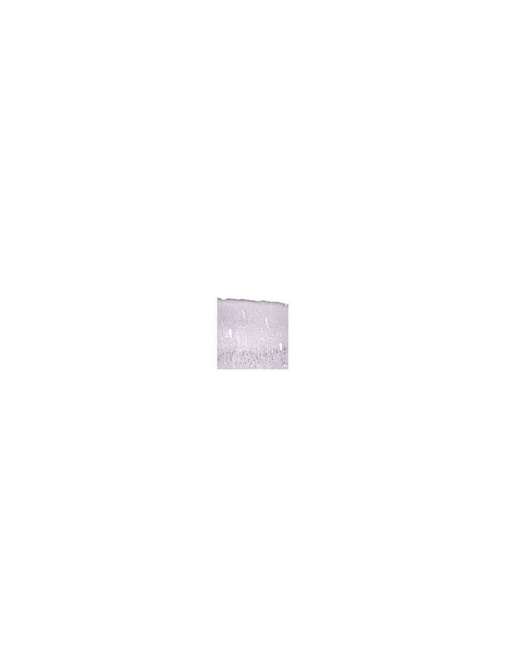
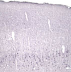
Immunohistochemical staining of Neurotrophin 4/5 (NT4/5) in rat cortex using rabbit polyclonal to recombinant human NT4, catalogue number R-102-500
Immunohistochemical staining of Neurotrophin 4/5 (NT4/5) in rat cortex using rabbit polyclonal to recombinant human NT4, catalogue number R-102-500
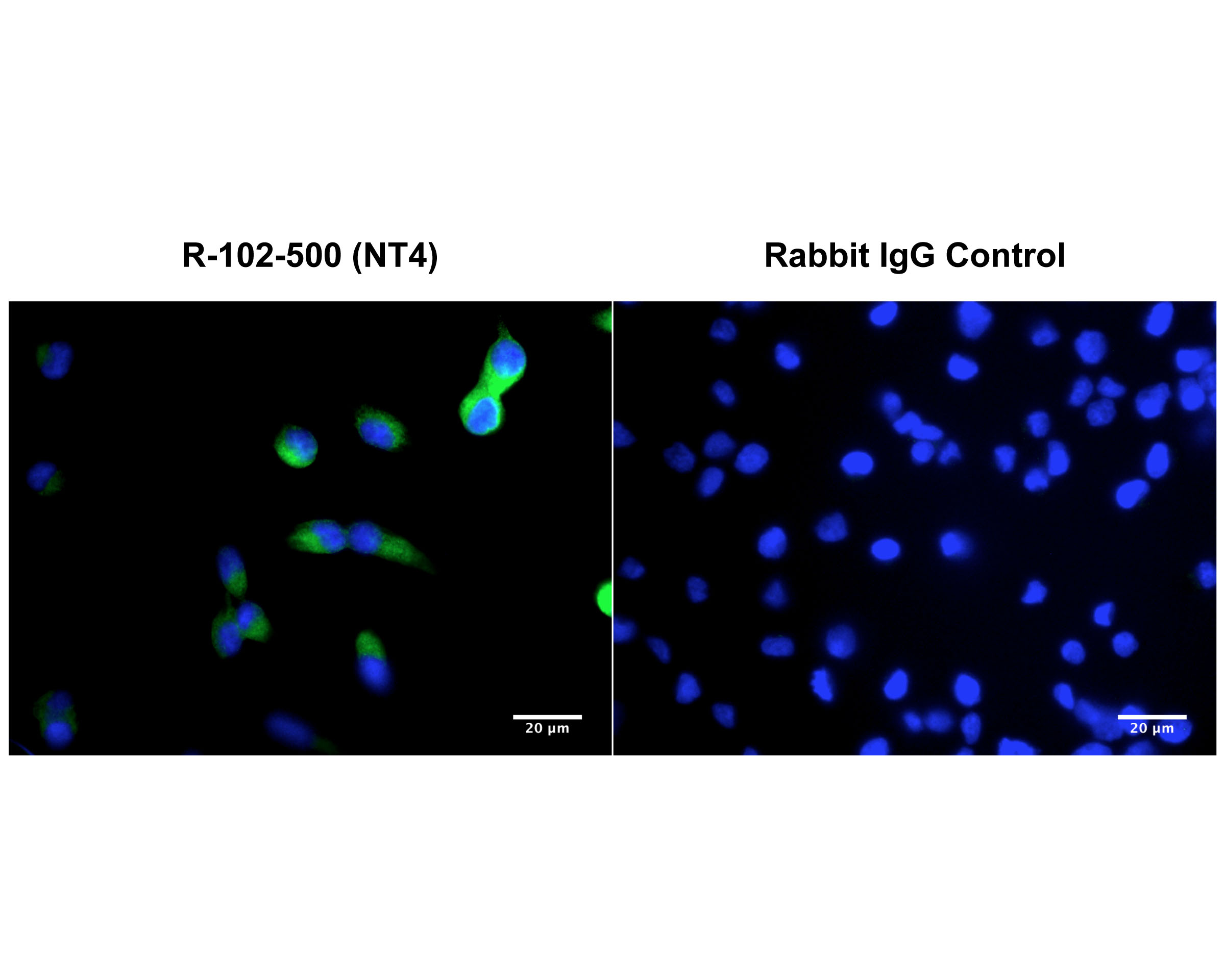
Immunofluorescence analysis of NT4 expression in human DU145 prostate cancer cells. Fixed (4% formaldehyde), permeabilized, and blocked (10% normal horse serum, 0.1% Triton X100) DU145 cells were incubated with NT4 rabbit antibody antibody R-102-500 (10 µg/mL, green) for 1 hour. Primary antibody binding was visualized with a secondary donkey anti-rabbit-CF488A antibody (4 µg/mL, 1 hour incubation). Control cells were incubated with normal rabbit IgG (10 µg/mL) as primary antibody. Cell nuclei were stained with Hoechst dye (blue). Magnification: 100x.
 1800 605-5127
 +61 (0)8 8352 7711