 1800 605-5127
 +61 (0)8 8352 7711
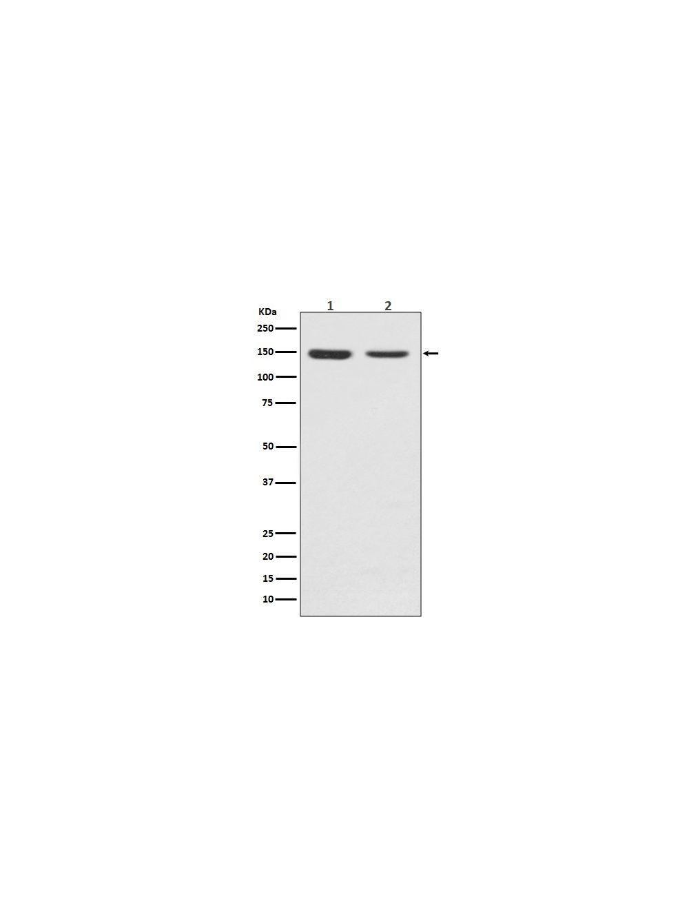

FGF Receptor 2, Rabbit Monoclonal Antibody
As low as
US$427.00
Only %1 left
Catalog Number
R-1776
- Product Name FGF Receptor 2, Rabbit Monoclonal Antibody
- Product Description Rabbit anti-FGF Receptor 2 Monoclonal Antibody (Unconjugated), suitable for WB, IP.
- Alternative Names FGFR-2; EC 2.7.10.1; Keratinocyte growth factor receptor 2; CD332; FGFR2; BEK; KGFR; KSAM;
- Application(s) IP, WB
- Antibody Host Rabbit
- Antibody Type Monoclonal
- Specificity The specificity of this antibody has been confirmed by WB on C6 cell lysates. The screened epitope of this antibody recognizes the protein of 135KD only, and the antibody has been verified via gene knockout.
- Species Reactivity Human, Mouse, Rat
- Immunogen Description A synthetic peptide from human FGF receptor 2.
- Conjugate Unconjugated
- Purity Description Rabbit IgG was purified from cell culture via protein G affinity chromatography.
- Regulatory Status For research use only.
Product Info
- Product Description Rabbit anti-FGF Receptor 2 Monoclonal Antibody (Unconjugated), suitable for WB, IP.
- Application(s) IP, WB
- Application Details Western Blotting (WB). A dilution of 1:500-1:2,000 is recommended for WB. R-1776-100 has an epitope located in the extracellular domain portion of FGFR2. In tests using C6 cells, this antibody detects a 135 kDa band under reducing conditions Immunoprecipitation (IP). A concentration of 1:50 is recommended for IP in a lysis buffer that contains no more than 0.5% detergent as well as proteinase inhibitors. Detection can be via R-1776-100 in westerns following, IP enrichment however rabbit heavy chain (50-55~ kDa) and light chains (~23-25 kDa) will also be detected by the anti-rabbit used to detect R-1776-100 on westerns unless biotinylation of R-1776-100 is used. As always, Biosensis recommends optimal dilutions/concentrations should be determined by the end user for their specific experimental design.
- Target FGF Receptor 2
- Specificity The specificity of this antibody has been confirmed by WB on C6 cell lysates. The screened epitope of this antibody recognizes the protein of 135KD only, and the antibody has been verified via gene knockout.
- Target Host Species Human
- Species Reactivity Human, Mouse, Rat
- Antibody Host Rabbit
- Antibody Type Monoclonal
- Antibody Isotype IgG
- Conjugate Unconjugated
- Immunogen Description A synthetic peptide from human FGF receptor 2.
- Purity Description Rabbit IgG was purified from cell culture via protein G affinity chromatography.
- Format Liquid in PBS pH 7.4, 150 mM NaCl, 0.02% sodium azide, 0.4-0.5 mg/mL BSA, 50% glycerol.
- Storage Instructions Maintain at -20°C unopened for up to 12 months after date of receipt. After opening maintain at -20°C in undiluted aliquots for up to six months.
- Batch Number Please see item label.
- Expiration Date 12 months after date of receipt (unopened vial).
- Alternative Names FGFR-2; EC 2.7.10.1; Keratinocyte growth factor receptor 2; CD332; FGFR2; BEK; KGFR; KSAM;
- Uniprot Number P21802
- Uniprot Number/Name P21802 (FGFR2_HUMAN)
- Scientific Background FGF receptor 2 (FGFR2) is a member of the fibroblast growth factor receptor family and is Single-pass type I membrane protein and a high-affinity receptor for acidic, basic and/or keratinocyte growth factor, depending on the isoform. At least 20 isoforms are produced by alternative splicing. Because of its many isoforms, FGF2 receptor can demonstrate a wide range of sizes on reduced westerns generally ranging from ~135 kDa to 50 kDa in size. FGFR2 is highly expressed in developing human tissues and localizes to plasma, Golgi, and cytoplasmic vesicles. After ligand binding, the activated receptor is rapidly internalized and degraded.
- Shipping Temperature 2-8°C (on cold packs)
- UNSPSC CODE 41116161
- Regulatory Status For research use only.
