Product NameTAR DNA-binding protein 43 (TDP-43), Mouse Monoclonal Antibody
Product DescriptiongoogleMouse anti-TAR DNA-binding protein 43 (TDP-43) Monoclonal Antibody (Unconjugated), suitable for WB, ICC, IHC-Frozen.
Alternative NamesTAR DNA-binding protein 43; TDP-43; TARDBP; TDP43;
Application(s)ICC, IHC-Frozen, WB
Antibody HostMouse
Antibody TypeMonoclonal
SpecificityThe specificity of this antibody has been confirmed by WB. This antibody detects ~43 kDa TDP43 protein on crude extract of mouse brain nuclear fraction. Human and Rodent. Predicted to react with other mammalian tissue.
Species ReactivityHuman, Mouse, Other Mammals (Predicted), Rat
Immunogen DescriptionThis antibody was raised against recombinant full length human his-tagged TDP43 which was expressed in E. coli and purified by nickel affinity.
Product DescriptionMouse anti-TAR DNA-binding protein 43 (TDP-43) Monoclonal Antibody (Unconjugated), suitable for WB, ICC, IHC-Frozen.
Application(s)ICC, IHC-Frozen, WB
Application DetailsWestern Blotting (WB), Immunocytochemistry (ICC) and Immunohistochemistry (IHC). A dilution of 1:1,000 - 1:5,000 is recommended for WB and IHC. A dilution of 1:500-1,000 is recommended for IC. Biosensis recommends optimal dilutions/concentrations should be determined by the end user.
TargetTAR DNA-binding protein 43 (TDP-43)
SpecificityThe specificity of this antibody has been confirmed by WB. This antibody detects ~43 kDa TDP43 protein on crude extract of mouse brain nuclear fraction. Human and Rodent. Predicted to react with other mammalian tissue.
Target Host SpeciesHuman
Species ReactivityHuman, Mouse, Other Mammals (Predicted), Rat
Antibody HostMouse
Antibody TypeMonoclonal
Antibody IsotypeIgG
Clone Name3H8
ConjugateUnconjugated
Immunogen DescriptionThis antibody was raised against recombinant full length human his-tagged TDP43 which was expressed in E. coli and purified by nickel affinity.
Purity DescriptionProtein G purified
FormatLyophilized from PBS buffer pH 7.2-7.6 with 0.1% trehalose, and sodium azide
Reconstitution InstructionsSpin vial briefly before opening. Reconstitute with 100 µL sterile-filtered, ultrapure water to achieve a 1 mg/mL concentration. Centrifuge to remove any insoluble material.
Storage InstructionsAfter reconstitution of lyophilized antibody, aliquot and store at -20°C for a higher stability. Avoid freeze-thaw cycles.
Batch NumberPlease see item label.
Expiration Date12 months after date of receipt (unopened vial).
Alternative NamesTAR DNA-binding protein 43; TDP-43; TARDBP; TDP43;
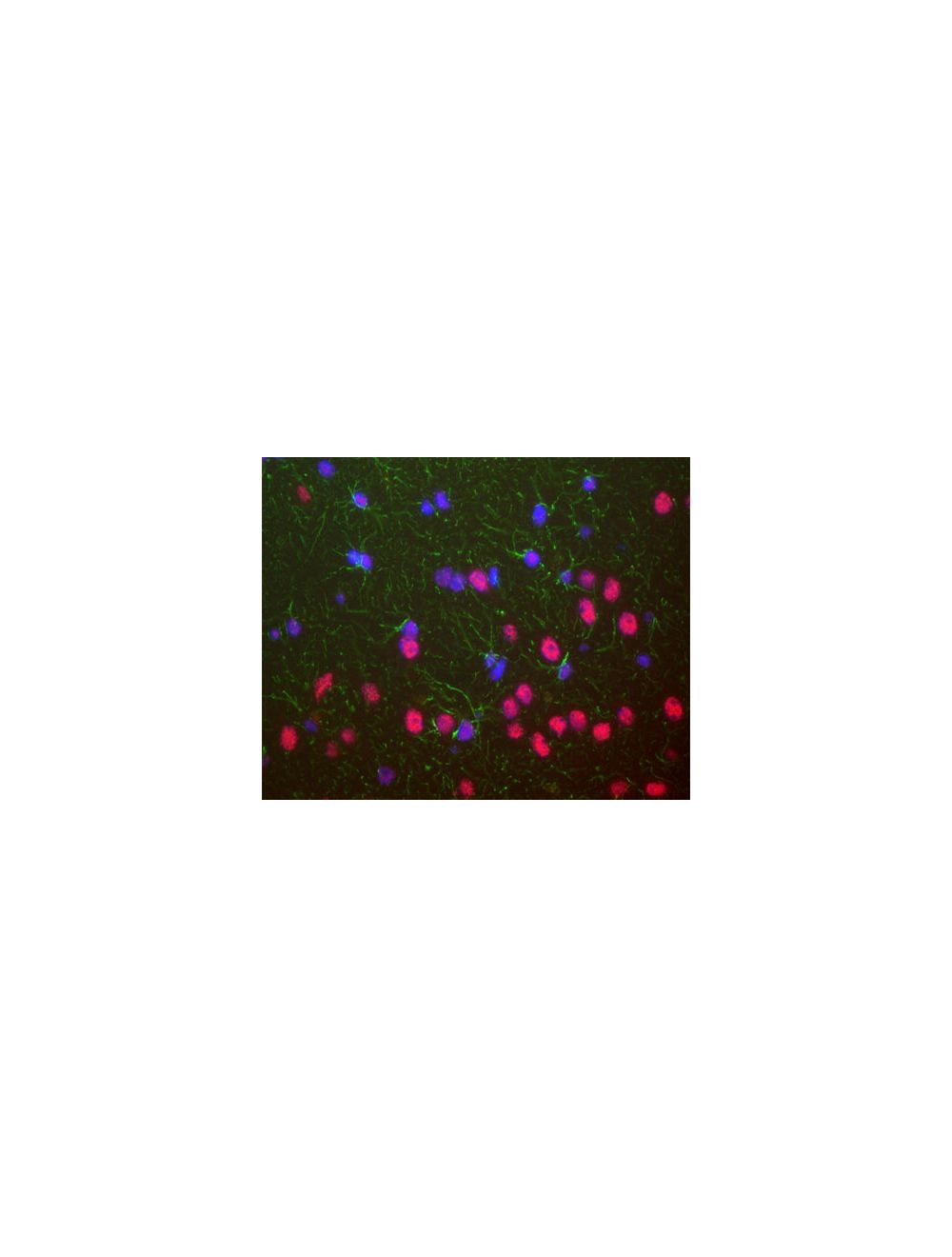
Mouse monoclonal antibody to TAR DNA-binding protein 43 [3H8] M-1403-100 was used to stain a section of formalin fixed adult rat brain, specifically the hippocampus. Hippocampal neuron nuclei are stained strongly. Chicken polyclonal antibody to GFAP C-1373-50 (green) shows the processes of astrocytic glial cells. Nuclei of all cells are revealed with DAPI DNA stain (blue). The TAR DNA-binding protein 43 antibody stains neuronal nuclei strongly and the nuclei of some non-neuronal cells much more weakly. Neuronal nuclei therefore look crimson, since they are both red due to the content of TAR DNA-binding protein 43 and blue due to their content of DNA, stained blue with DAPI.
Left: Analysis of TDP43 expression in rat hippocampus section by Immunohistochemistry. Section was stained with mouse anti-TDP43 antibody (red, 1:2,000), and co-stained with chicken antibody to GFAP (C-1373-50, green, 1:5,000). Blue: DAPI nuclear stain. IHC method: Following transcardial perfusion of rat with 4% paraformaldehyde, brain was post-fixed for 24 hours, cut to 45 um sections, and free-floating sections were stained. The TDP43 protein is concentrated in the nuclei of hippocampal neurons, while the GFAP antibody stains the intermediate filament network of astroglial cells. Right: Western blot analysis of whole brain lysates and nuclear extracts for TDP43 expression (green, 1:2,000). [1] protein standard, [2] rat brain, [3] rat brain nuclear extract, [4] mouse brain, [5] mouse brain nuclear extract. A strong band at 43 kDa corresponds to TDP43 protein.
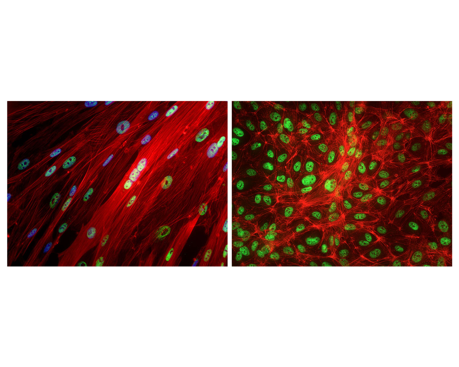
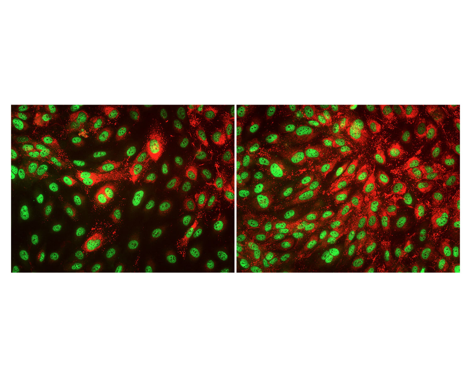
TDP43 staining in fixed and permeabilized human umbilical vein endothelial cells (HUVEC) by Immunocytochemistry. TDP43-IR (green) is localized to cell nuclei as expected. Blue: Hoechst nuclear stain (left only). Red: Phalloidin staining. Primary TDP43 antibody concentration: 2 µg/mL; Magnification: 32X. Image courtesy of QBM Cell Science.
TDP43 staining in fixed and permeabilized human umbilical vein endothelial cells (HUVEC) by Immunocytochemistry. TDP43-IR (green) is localized to cell nuclei as expected. Red: Staining of von Willebrandt factor. Primary TDP43 antibody concentration: 2 µg/mL; Magnification: 32X. Image courtesy of QBM Cell Science.
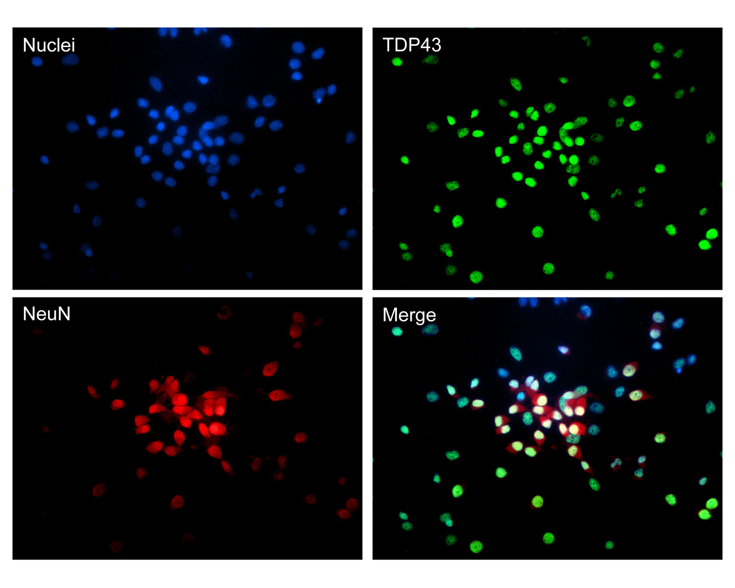
Staining of fixed and permeabilized rat hippocampal cells for TDP43 (green) and NeuN (red) by Immunocytochemistry. Green: Mouse antibody to TDP43, M-1403-100 (2 µg/mL); Red: Rabbit antibody to NeuN, R-3770-100 (1:500 dilution); Blue: Hoechst nuclear dye. TDP43-IR and NeuN-IR is localized to cell nuclei as expected. Magnification: 32X. Image courtesy of QBM Cell Science.
 1800 605-5127
 +61 (0)8 8352 7711