 1800 605-5127
 +61 (0)8 8352 7711
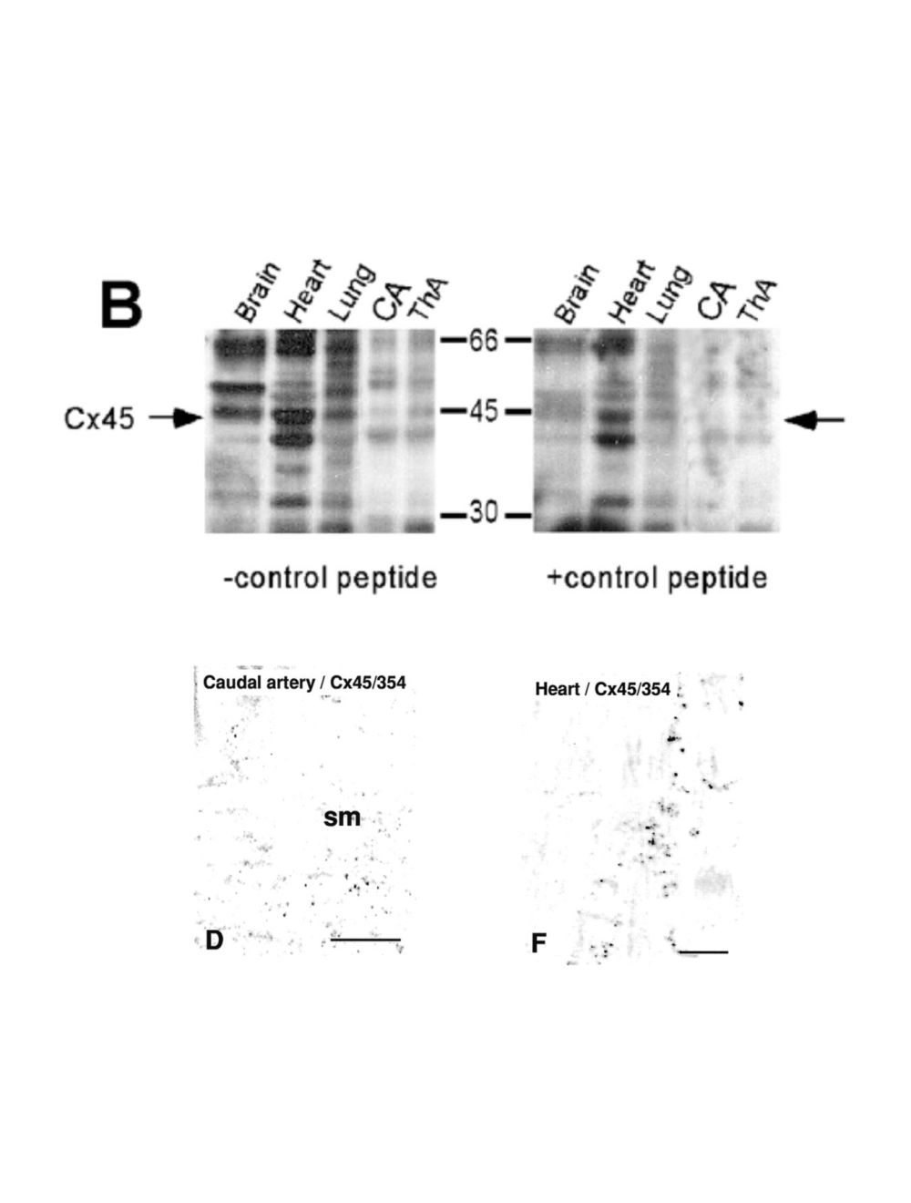

Connexin-45 (Cx45), Sheep Polyclonal Antibody
As low as
US$327.00
Only %1 left
Catalog Number
S-061
- Product Name Connexin-45 (Cx45), Sheep Polyclonal Antibody
- Product Description Sheep anti-Connexin-45 (Cx45) Polyclonal Antibody (Unconjugated), suitable for WB.
- Alternative Names Gap junction gamma-1 protein; Gap junction alpha-7 protein; Cx45; GJC1; GJA7
- Application(s) WB
- Antibody Host Sheep
- Antibody Type Polyclonal
- Specificity Western blotting of cell membranes of COS cells transiently transfected with cDNA encoding Connexin-45 shows a singular band of molecular weight 47 kDa which is abolished by preincubation with the immunising peptide. No such band is detected in western blots of membranes from non-transfected cells or cells transfected with cDNA encoding Connexin-37 or Connexin-40. This antiserum reacts to human Connexin-45.
- Species Reactivity Human
- Immunogen Description A synthetic peptide (QAYSHQN NPHGPRE) as part of human Connexin-45 protein (aa: 354-367) conjugated to diphtheria toxoid has been used as the immunogen.
- Conjugate Unconjugated
- Purity Description Whole serum
- Regulatory Status For research use only.
Product Info
- Product Description Sheep anti-Connexin-45 (Cx45) Polyclonal Antibody (Unconjugated), suitable for WB.
- Application(s) WB
-
Application Details
Immunohistochemistry: Antibody detects sparse Cxn45-IR in caudal artery and heart tissue (see Rummery, NM et al 2002 for more staining specifics). Antibody was used at 1:100 to 1:250, but Biosensis recommends optimal dilutions/concentrations should be determined by the end user. In the original work the specificity of the antibody was shown by incubation either without primary antibody or with primary antibody that had previously been pre-incubated for 1 hour at room temperature with 10-fold excess by weight of the peptide against which the antibody was raised.
Western Blot: Antibody is not recommended for western blotting by Biosensis, however, it does react in westerns with Cxn 45 specific material. The authors report that the antibody develops numerous bands in westerns blots, only some of which are removed upon peptide treatment (see Rummery, NM et al 2002). The Cx45/354 antibody revealed the presence of a specific 45-kDa band in all tissues tested, although it was very weak in the arteries. A higher molecular weight band, which was blocked by peptide, was also seen in the brain (see Rummery, NM et al 2002, online Figure VIIB, -/+ peptide). - Target Connexin-45 (Cx45)
- Specificity Western blotting of cell membranes of COS cells transiently transfected with cDNA encoding Connexin-45 shows a singular band of molecular weight 47 kDa which is abolished by preincubation with the immunising peptide. No such band is detected in western blots of membranes from non-transfected cells or cells transfected with cDNA encoding Connexin-37 or Connexin-40. This antiserum reacts to human Connexin-45.
- Target Host Species Human
- Species Reactivity Human
- Antibody Host Sheep
- Antibody Type Polyclonal
- Antibody Isotype Mixed
- Conjugate Unconjugated
- Immunogen Description A synthetic peptide (QAYSHQN NPHGPRE) as part of human Connexin-45 protein (aa: 354-367) conjugated to diphtheria toxoid has been used as the immunogen.
- Sequence QAYSHQNNPHGPRE
- Purity Description Whole serum
- Format Lyophilized
- Reconstitution Instructions Spin vial briefly before opening. Reconstitute in 100 µL sterile-filtered, ultrapure water. Centrifuge to remove any insoluble material.
- Storage Instructions After reconstitution keep aliquots at -20°C for a higher stability, and at 2-8°C with an appropriate antibacterial agent. Glycerol (1:1) may be added for an additional stability. Avoid repetitive freeze/thaw cycles.
- Batch Number Please see item label.
- Expiration Date 12 months after date of receipt (unopened vial).
- Alternative Names Gap junction gamma-1 protein; Gap junction alpha-7 protein; Cx45; GJC1; GJA7
- Uniprot Number P36383
- Uniprot Number/Name P36383 (CXG1_HUMAN)
- Scientific Background Connexin-45 is a component of gap junctions, which are composed of a cluster of closely packed pairs of transmembrane channels, the connexons, through which materials of low molecular weight diffuse from one cell to a neighboring cell. SUBUNIT: A connexon is composed of a hexamer of connexins. SUBCELLULAR LOCATION: Membrane; multi-pass membrane protein. SIMILARITY: Belongs to the connexin family. Alpha-type (group II) subfamily. Alternatively spliced isoforms have been described.
- Shipping Temperature 25°C (ambient)
- UNSPSC CODE 41116161
- Regulatory Status For research use only.
Specifications

-
Specific References
Original Reference
Rummery NM, Hickey H, McGurk G, Hill CE. (2002) "Connexin37 is the major connexin expressed in the media of caudal artery.” Arterioscler Thromb Vasc Biol. 22(9):1427-32. PMID: 12231561
This antibody is referred to as Cx45/354 in Rummery, NM et al 2002.