Alternative NamesSARS-CoV-2 (COVID-19) Spike Antibody: Severe acute respiratory syndrome coronavirus 2 (SARS-CoV-2), Surface Glycoprotein, Spike protein
Application(s)ELISA, ICC, WB
Antibody HostRabbit
Antibody TypePolyclonal
Specificity Predicted reactivity based on immunogen sequence: SARS-CoV Spike proteins: (100%)
Species ReactivityVirus
Immunogen DescriptionAnti-SARS-CoV-2 (COVID-19) Spike antibody (R-1953-100) was raised against a peptide corresponding to 20 amino acids near the carboxy terminus of SARS-CoV-2 (COVID-19) Spike glycoprotein.
The immunogen is located within the last 50 amino acids of SARS-CoV-2 (COVID-19) Spike protein.
ConjugateUnconjugated
Concentration1 mg/mL
Purity DescriptionAffinity-purified via peptide column.
Antibody validated: Immunofluorescence in human samples. SARS-CoV-2 (COVID-19) Spike antibody can be used for the detection of SARS-CoV-2 (COVID-19) Spike protein in ELISA. It will detect 4 ng of free peptide at 1 μg/mL. All other applications and species not yet tested.
TargetSARS-CoV-2 surface glycoprotein
Specificity Predicted reactivity based on immunogen sequence: SARS-CoV Spike proteins: (100%)
Target Host SpeciesVirus
Species ReactivityVirus
Antibody HostRabbit
Antibody TypePolyclonal
Antibody IsotypeIgG
ConjugateUnconjugated
Immunogen DescriptionAnti-SARS-CoV-2 (COVID-19) Spike antibody (R-1953-100) was raised against a peptide corresponding to 20 amino acids near the carboxy terminus of SARS-CoV-2 (COVID-19) Spike glycoprotein.
The immunogen is located within the last 50 amino acids of SARS-CoV-2 (COVID-19) Spike protein.
HomologyPredicted reactivity based on immunogen sequence: SARS-CoV Spike proteins: (100%)
Isoform InformationSARS-CoV-2 (COVID-19) Spike has one isoform (1273aa).
Purity DescriptionAffinity-purified via peptide column.
FormatLiquid. SARS-CoV-2 (COVID-19) Spike antibody is supplied in PBS containing 0.02% sodium azide. Conc.1 mg/mL
Concentration1 mg/mL
Reconstitution InstructionsSpin vial briefly before opening.
Storage InstructionsSARS-CoV-2 (COVID-19) Spike antibody can be stored at 2-8°C for three months and -20°C, stable for up to one year. As with all antibodies care should be taken to avoid repeated freeze thaw cycles. Antibodies should not be exposed to prolonged high temperatures.
Batch NumberPlease see item label.
Expiration Date12 months after date of receipt (unopened vial).
Alternative NamesSARS-CoV-2 (COVID-19) Spike Antibody: Severe acute respiratory syndrome coronavirus 2 (SARS-CoV-2), Surface Glycoprotein, Spike protein
Scientific BackgroundCoronavirus disease 2019 (COVID-19), formerly known as 2019-nCoV acute respiratory disease, is an infectious disease caused by SARS-CoV-2, a virus closely related to the SARS virus (1). The disease is the cause of the 2019–20 coronavirus outbreak (2). The structure of 2019-nCoV consists of the following: a Spike protein (S), hemagglutinin-esterease dimer (HE), a membrane glycoprotein (M), an envelope protein (E) a nucleoclapid protein (N) and RNA. Coronavirus invades cells through Spike (S) glycoproteins, a class I fusion protein. It is the major viral surface protein that coronavirus uses to bind to the human cell surface receptor. It also mediates the fusion of host and viral cell membrane, allowing the virus to enter human cells and begin infection (3). The spike protein is the major target for neutralizing antibodies and vaccine development (4). The protein modeling suggests that there is strong interaction between Spike protein receptor-binding domain and its host receptor angiotensin-converting enzyme 2 (ACE2), which regulate both the cross-species and human-to-human transmissions of COVID-19 (5). The recent study has shown that the SARS-CoV-2 spike protein binds ACE2 with higher affinity than SARS-CoV spike protein (6).
Figure 1 Overexpression Validation in Spike Transfected 293 Cells Loading: 15 μg per lane of 293 cell lysate. Antibodies: SARS-CoV-2 (COVID-19) Spike, R-1953-100 (1 μg/mL), 1h incubation at RT in 5% NFDM/TBST. Secondary: Goat anti-rabbit IgG HRP conjugate at 1:10000 dilution. Lane 1: WT 293 cells and Lane 2: SARS-CoV-2 Spike overexpressed 293 cells
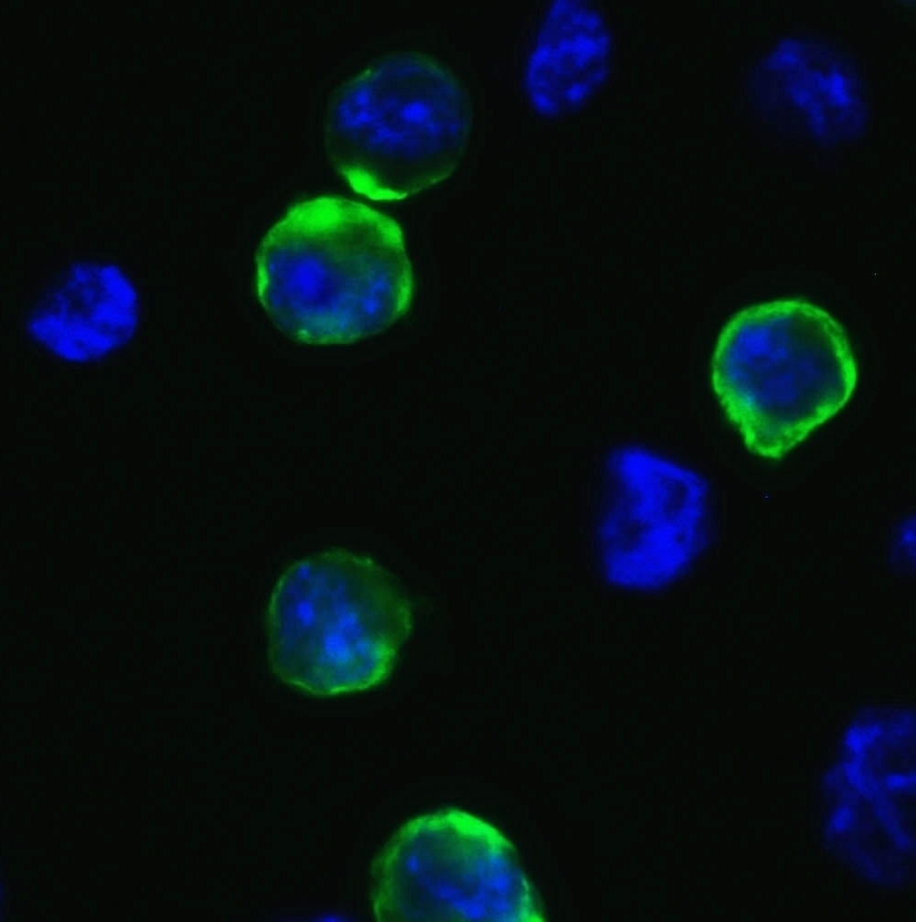
Figure 2 Immunofluorescence Validation of SARS-CoV-2 (COVID-19) Spike in 293 Transfected Cells Immunofluorescent analysis of 4% paraformaldehyde-fixed 293 spike transfected cells labeling SARS-CoV-2 (COVID-19) Spike with R-1953-100 at 1 μg/mL, followed by goat anti-rabbit IgG secondary antibody at 1/500 dilution (green) and DAPI staining (blue).
Figure 3 ELISA Test Antibodies: SARS-CoV-2 (COVID-19) Spike antibody, R-1953-100 (1 μg/mL). A direct ELISA was performed using immunogen or control peptide as coating antigen and the anti-SARS-CoV-2 (COVID-19) Spike antibody as the capture antibody. Secondary: Goat anti-rabbit IgG HRP conjugate at 1:20000 dilution. Detection range is from 0.5 ng/mL to 1000ng/mL.
General ReferencesGorbalenya. bioRxiv: 2020. Hui et al. Int J Infect Dis. 2020;91:264-266. Belouzard et al. Viruses. 2012;4(6):1011-33. Lee et al. J Virol. 2006;80(8):4079-87. Wan et al. J Virol. 2020. Wrapp et al. Science. 2020.
 1800 605-5127
 +61 (0)8 8352 7711