Product NameTyrosine Kinase Receptor A (TrkA), Mouse Monoclonal Antibody
Product DescriptiongoogleMouse anti-Tyrosine Kinase Receptor A (TrkA) Monoclonal Antibody (Unconjugated), suitable for WB, ICC.
Alternative NamesTropomyosin-related kinase receptor; High affinity nerve growth factor receptor; Neurotrophic tyrosine kinase receptor type 1; TRK1 transforming tyrosine kinase protein; p140-TrkA; Trk-A; NTRK1; TRK
Application(s)ICC, WB
Antibody HostMouse
Antibody TypeMonoclonal
SpecificityTrkA. Does not cross-react with TrkB or TrkC. Reacts with human TrkA. Known to cross-react with TrkA from rat and mouse. Expected to cross-react with other mammalian species based on peptide antigen sequence similarity.
Species ReactivityHuman, Mouse, Other Mammals (Predicted), Rat
Immunogen DescriptionA synthetic peptide from the intracellular cytoplasmic domain of human TrkA (C-HGPDAKLLAGGE, aa: 604-615) has been used as the immunogen.
ConjugateUnconjugated
Purity DescriptionProtein G purified mouse immunoglobulin
Product DescriptionMouse anti-Tyrosine Kinase Receptor A (TrkA) Monoclonal Antibody (Unconjugated), suitable for WB, ICC.
Application(s)ICC, WB
Application DetailsWB: Western blotting: 1-3 µg/mL, antibody is suitable for reduced (with DTT or beta-mercaptoethanol) and non-reduced samples. Denatured but not reduced samples provides cleaner blot signals as demonstrated in our photographs.
Immunocytochemistry: 1-2 µg/mL. Antibody works on 4% formaldehyde-fixed cells. Note that cells require a permeabilization step, because the antibody detects a cytoplasmic epitope of TrkA.
Other applications not yet tested. Biosensis recommends optimal dilutions/concentrations should be determined by the end user.
TargetTyrosine Kinase Receptor A (TrkA)
SpecificityTrkA. Does not cross-react with TrkB or TrkC. Reacts with human TrkA. Known to cross-react with TrkA from rat and mouse. Expected to cross-react with other mammalian species based on peptide antigen sequence similarity.
Target Host SpeciesHuman
Species ReactivityHuman, Mouse, Other Mammals (Predicted), Rat
Antibody HostMouse
Antibody TypeMonoclonal
Antibody IsotypeIgG3, kappa
Clone NameBS292
ConjugateUnconjugated
Immunogen DescriptionA synthetic peptide from the intracellular cytoplasmic domain of human TrkA (C-HGPDAKLLAGGE, aa: 604-615) has been used as the immunogen.
SequenceHGPDAKLLAGGE
Purity DescriptionProtein G purified mouse immunoglobulin
FormatLyophilized from PBS, pH 7.4, containing 3% trehalose without preservatives.
Reconstitution InstructionsSpin vial briefly before opening. Reconstitute in 100 µL sterile-filtered, ultrapure water. Centrifuge to remove any insoluble material. Final buffer contains no preservatives.
Storage InstructionsAfter reconstitution divide into aliquots and store at -20°C for a higher stability. Antibody contains no preservatives. Store at 2-8°C with an appropriate antibacterial agent. Use sterile methods. Highest purity Glycerol (1:1) may be added for an additional stability when stored at refrigerated or freezing temperatures. Avoid repetitive freeze/thaw cycles.
Batch NumberPlease see item label.
Expiration Date12 months after date of receipt (unopened vial).
Alternative NamesTropomyosin-related kinase receptor; High affinity nerve growth factor receptor; Neurotrophic tyrosine kinase receptor type 1; TRK1 transforming tyrosine kinase protein; p140-TrkA; Trk-A; NTRK1; TRK
Scientific BackgroundTrkA is a member of the neurotrophic tyrosine kinase receptor family. It is a membrane-bound receptor that upon neurotrophin binding, phosphorylates itself and members of the MAPK pathway. TrkA is required for high-affinity binding to nerve growth factor (NGF), neurotrophin-3 and neurotrophin-4/5 but not brain-derived neurotrophic factor (BDNF). TrkA leads to cell differentiations and may play a role in specifying sensory neuron subtypes. It has a crucial role in the development and function of the nociceptive reception system as well as establishment of thermal regulation via sweating. SUBUNIT: Exists in a dynamic equilibrium between monomeric (low affinity) and dimeric (high affinity) structures. SUBCELLULAR LOCATION: Cell membrane; single-pass type I membrane protein. Endocytosed to the endosomes upon treatment of cells with NGF. ALTERNATIVE PRODUCTS: 2 named isoforms produced by alternative splicing. Both isoforms have similar biological properties. TISSUE SPECIFICITY: Isoform TrkA-II is primarily expressed in neuronal cells. Isoform TrkA-I is found in non-neuronal tissues. Mutations in TrkA have been associated with congenital insensitivity to pain, anhidrosis, self-mutalating behaviour, mental retardation and cancer (Reference: www.uniprot.com).
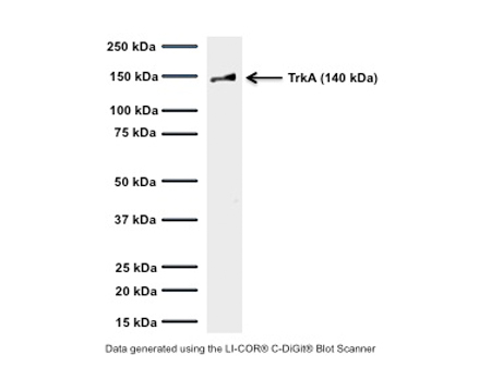
Western blot of TrkA in rat PC12 cell lysates (64 µg/lane): SDS-PAGE: denatured, non-reduced; Transfer: Tris-Glycine buffer; Membrane: nitrocellulose (0.45 um); Blocking: 5% skim milk in TBST, 1 hour at RT; Primary antibody: overnight at 4°C (3 µg/mL); Secondary antibody: anti-mouse-HRP (1/6000) 2 hours at RT; Detection: Chemiluminiscence. M-1719-100 detects membrane-associated TrkA full-length protein at approximately 140 kDa (gp140TrkA). Predicted MW of rat TrkA based on amino acid sequence: 85 kDa. The observed MW differs due to post-translational modification, mainly glycosylation.
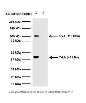
Western blot of TrkA in rat PC12 cell lysates (80 µg/lane). M-1719-100 detects the intracellular glycosylated immature form of TrkA at 110 kDa (gp140TrkA), and an intracellular cleavage product at 41 kDa (p41, see Cabrera et al., 1995; Rossi et al., 2002). These bands are TrkA-specific, because detection by M-1719-100 is abolished after pre-incubation with blocking peptide. SDS-PAGE: denatured, non-reduced; Transfer: Tris-Glycine buffer; Membrane: nitrocellulose (0.45 µm); Blocking: 5% skim milk in TBST, 1 hour at RT; Primary antibody: overnight at 4°C (3 µg/mL); Secondary antibody: anti-mouse-HRP (1/6000) 2 hours at RT; Detection: Chemiluminiscence.
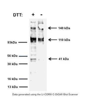
Western blot of TrkA in rat PC12 cell lysates (30 µg/lane). M-1719-100 detects membrane-associated TrkA full-length protein at 140 kDa (gp140TrkA), the glycosylated immature form of TrkA at 110 kDa (gp110TrkA) and an intracellular cleavage product at 41 kDa (p41, see Cabrera et al., 1995; Rossi et al., 2002). SDS-PAGE: denatured, reduced and non-reduced; Transfer: Tris-Glycine buffer; Membrane: nitrocellulose (0.45 µm); Blocking: 5% skim milk in TBST, 2 hours at RT; Primary antibody: overnight at 4°C (3 µg/mL); Secondary antibody: anti-mouse-HRP (1/6000) 2 hours at RT; Detection: Chemiluminiscence.
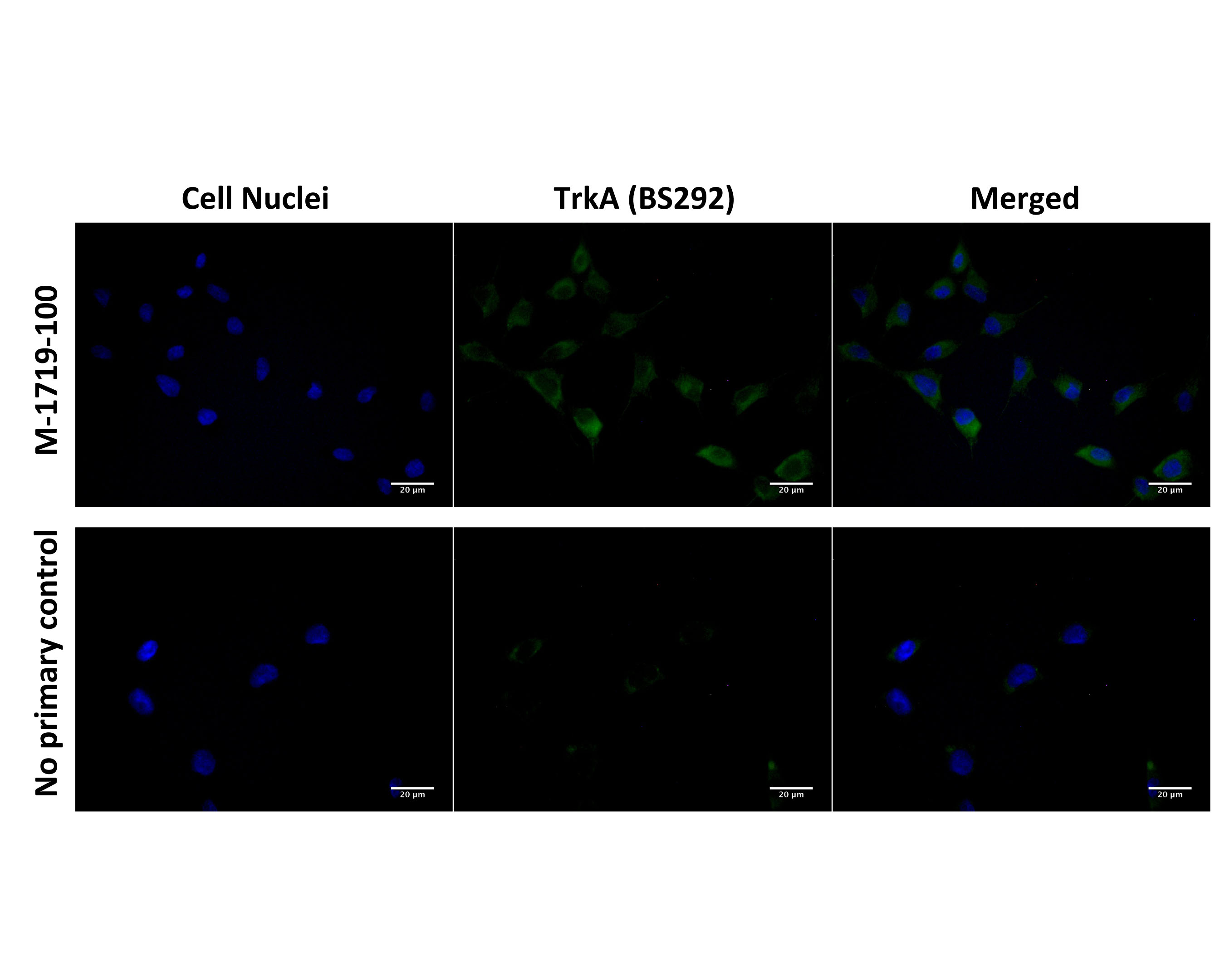
Immunofluorescence analysis of TrkA expression in human SH-SY5Y cells. Fixed (4% formaldehyde), permeabilized, and blocked (10% normal horse serum, 0.1% Triton X100) SH-SY5Y cells were incubated with TrkA antibody M-1719-100 (2 µg/mL, green) for 1 hour. Primary antibody binding was visualized with a secondary donkey anti-mouse-CF488A antibody (4 µg/mL, 1 hour incubation). Cell nuclei were stained with Hoechst dye (blue). Magnification: 100x.
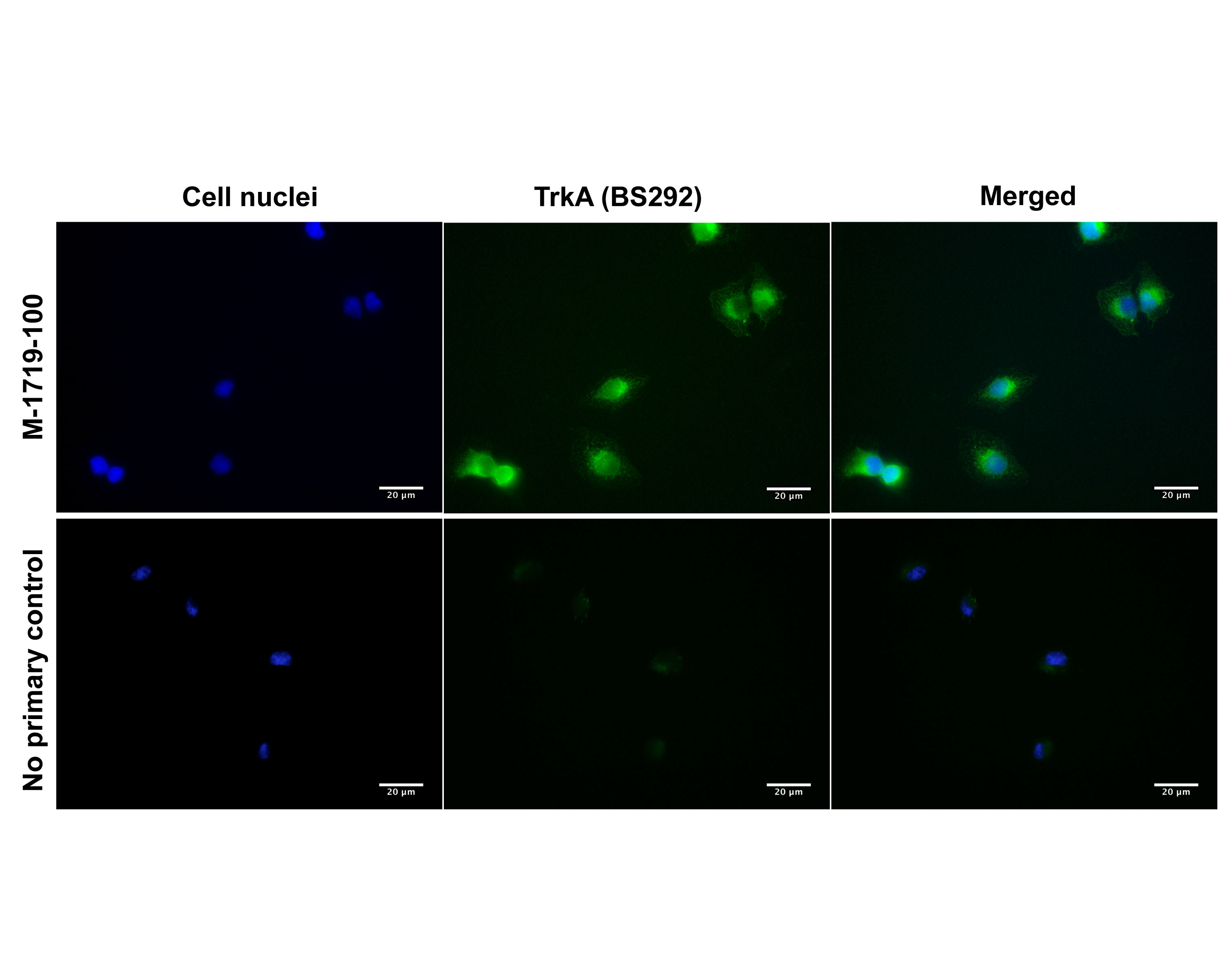
Immunofluorescence analysis of TrkA expression in human DU145 prostate cancer cells. Fixed (4% formaldehyde), permeabilized, and blocked (10% normal horse serum, 0.1% Triton X100) DU145 cells were incubated with TrkA antibody M-1719-100 (2 µg/mL, green) for 1 hour. Primary antibody binding was visualized with a secondary donkey anti-mouse-CF488A antibody (4 µg/mL, 1 hour incubation). Cell nuclei were stained with Hoechst dye (blue). Magnification: 100x.
TrkA staining in fixed and permeabilized embryonic rat DRG neurons by Immunocytochemistry. TrkA-IR is observed in axonal projections (white arrows) and cell body (membrane and/or cytoplasmic compartments, yellow arrow), consistent with expected cellular localization. Primary antibody concentration: 2 µg/mL; Magnification: 32X. Image courtesy of QBM Cell Science.
General ReferencesCabrera N. et al., TrkA receptor ectodomain cleavage generates a tyrosine-phosphorylated cell-associated frangment. J Cell Biol, 1996, Vol.132(3), pp.427-436. Rossi F.M. et al., Expression of the nerve growth factor receptors TrkA and p75NTR in the visual cortex of the rat: development and regulation by the cholinergic input. J Neurosci, Februar 1, 2002, 22(3):912-919.
 1800 605-5127
 +61 (0)8 8352 7711