 1800 605-5127
 +61 (0)8 8352 7711
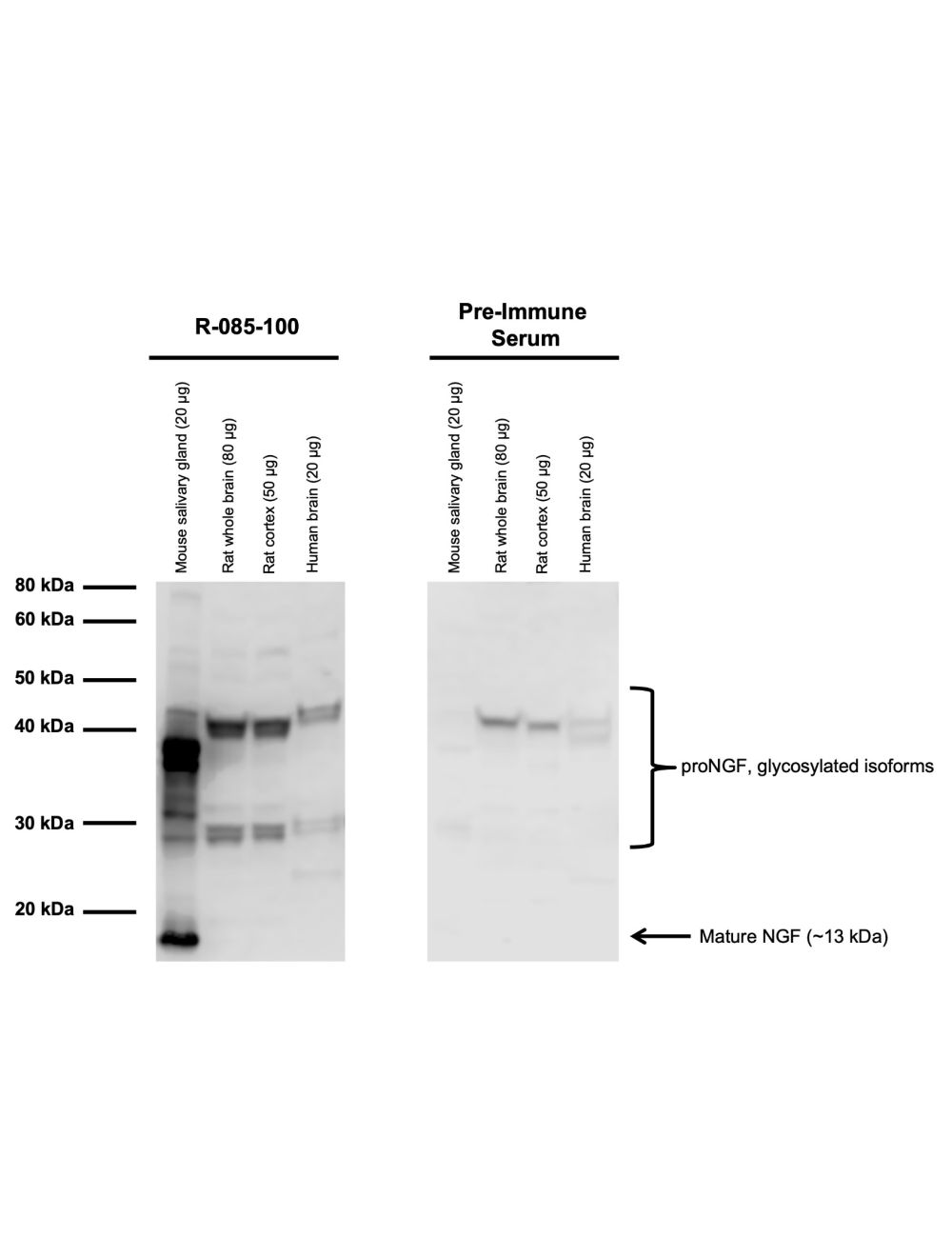

Beta-nerve growth factor (beta-NGF), Rabbit Polyclonal Antibody
- Product Name Beta-nerve growth factor (beta-NGF), Rabbit Polyclonal Antibody
- Product Description Rabbit anti-Beta-nerve growth factor (beta-NGF) Polyclonal Antibody (Unconjugated), suitable for WB, IHC-Frozen, ELISA, Neutralize.
- Alternative Names Beta-nerve growth factor
- Application(s) ELISA, IHC-Frozen, Neutralize, WB
- Antibody Host Rabbit
- Antibody Type Polyclonal
- Specificity A cross reactivity of less than 1% to recombinant human BDNF, NT3, NT4/5 by ELISA has been shown. This antiserum is known to cross react with mouse, rat, human and avian NGF bot not bovine NGF.
- Species Reactivity Avian, Human, Mouse, Rat
- Immunogen Description Native mouse beta NGF purified from submaxillary salivary gland (95% purity by PAGE)
- Conjugate Unconjugated
- Purity Description Whole serum
- Regulatory Status For research use only.
Product Info
- Product Description Rabbit anti-Beta-nerve growth factor (beta-NGF) Polyclonal Antibody (Unconjugated), suitable for WB, IHC-Frozen, ELISA, Neutralize.
-
Related Products
Nerve growth factor (2.5S), Mouse, Purified Native Protein
Beta-nerve growth factor (Beta-NGF), Rabbit Polyclonal Antibody
- Application(s) ELISA, IHC-Frozen, Neutralize, WB
- Application Details IHC, 1-site ELISA, WB, immunoblot, inhibition of biological activity. A dilution of 1:1000-1:5000 is recommended for IHC, western blot and immunoblot; 1:15000 for ELISA; for inhibition of biological activity: 1:10-50 for in vitro, 5-10 µL/g body weight for in vivo. This antiserum completely inhibits neuronal survival and the outgrowth actions of murine NGF in chicken DRG in vitro. Biosensis recommends optimal dilutions/concentrations should be determined by the end user.
- Target Beta-nerve growth factor (beta-NGF)
- Specificity A cross reactivity of less than 1% to recombinant human BDNF, NT3, NT4/5 by ELISA has been shown. This antiserum is known to cross react with mouse, rat, human and avian NGF bot not bovine NGF.
- Target Host Species Mouse
- Species Reactivity Avian, Human, Mouse, Rat
- Antibody Host Rabbit
- Antibody Type Polyclonal
- Antibody Isotype Mixed
- Conjugate Unconjugated
- Immunogen Description Native mouse beta NGF purified from submaxillary salivary gland (95% purity by PAGE)
- Purity Description Whole serum
- Format Lyophilized
- Reconstitution Instructions Spin vial briefly before opening. Reconstitute in 100 µL of sterile-filtered, ultrapure water. Centrifuge to remove any insoluble material.
- Storage Instructions Store lyophilized antibody at 2-8ºC. After reconstitution keep aliquots at -20°C to -80ºC for a higher stability, and at 2-8°C with an appropriate antibacterial agent. Avoid repetitive freeze/thaw cycles. Glycerol (1:1) may be added for an additional stability.
- Batch Number Please see item label.
- Expiration Date 12 months after date of receipt (unopened vial).
- Alternative Names Beta-nerve growth factor
- Uniprot Number P01139
- Uniprot Number/Name P01139 (NGF_MOUSE)
- Scientific Background FUNCTION: Nerve growth factor is important for the development and maintenance of the sympathetic and sensory nervous systems. It stimulates division and differentiation of sympathetic and embryonic sensory neurons. SUBUNIT: Homodimer, associated by noncovalent forces. SUBCELLULAR LOCATION: Secreted protein. SIMILARITY: Belongs to the NGF-beta family.
- Shipping Temperature 25°C (ambient)
- UNSPSC CODE 41116161
- Regulatory Status For research use only.
Specifications

- Specific References Mulhall J.P. et al (2008) J Sex Med. May;5(5):1126-36.
-
General References
Ebendal, T. et al (1989) J Neurosci Res 22, 223-240.
Zhou, X. F. et al (1994) J Neurosci Methods 54, 95-102.
Angeletti, P. U. et al (1968) Adv Enzymol Relat Areas Mol Biol 31, 51-7
Hesse K. et al. (1997) Neurosci Lett. Aug 8;231(2):83-
Miao J et al. (2012) Neurosci Res. Dec;74(3-4):269-7