Application DetailsImmunohistochemistry (IHC) and immunocytochemistry (ICC): 1-2 µg/mL. This antibody has been shown to work on 4% PFA fixed mouse brain sections.
Western blotting (WB): 0.5-1.0 µg/mL. This antibody detects bands between 50-65 kDa, which only appear in stimulated cells.
Biosensis recommends optimal dilutions/concentrations should be determined by the end user.
TargetCellular oncogene fos (c-Fos)
SpecificityHuman. Horse, cow, pig, chicken, rat, mouse.
Target Host SpeciesHuman
Species ReactivityBovine, Chicken, Horse, Human, Mouse, Pig, Rat
Antibody HostMouse
Antibody TypeMonoclonal
Antibody IsotypeIgG1
Clone Name2H2
ConjugateUnconjugated
Immunogen DescriptionFull length, E.coli-derived recombinant human c-FOS protein.
Purity DescriptionProtein G purified
FormatLyophilized from PBS buffer pH 7.2-7.6 with 0.1% trehalose, and sodium azide
Reconstitution InstructionsSpin vial briefly before opening. Reconstitute with 100 µL sterile-filtered, ultrapure water to achieve a 1 mg/mL concentration. Centrifuge to remove any insoluble material.
Storage InstructionsStore lyophilized, unopened vial at 2-8°C or lower. After reconstitution, prepare aliquots and store at -20°C for a higher stability. Avoid freeze-thaw cycles.
Batch NumberPlease see item label.
Expiration Date12 months after date of receipt (unopened vial).
Alternative NamesCellular oncogene fos; G0/G1 switch regulatory protein 7; cFOS
Scientific BackgroundFUNCTION: Nuclear phosphoprotein which forms a tight but non-covalently linked complex with the JUN/AP-1 transcription factor. In the heterodimer, FOS and JUN/AP-1 basic regions each seems to interact with symmetrical DNA half sites. On TGF-beta activation, forms a multimeric SMAD3/SMAD4/JUN/FOS complex at the AP1/SMAD-binding site to regulate TGF-beta-mediated signaling. Has a critical function in regulating the development of cells destined to form and maintain the skeleton. It is thought to have an important role in signal transduction, cell proliferation and differentiation. In growing cells, activates phospholipid synthesis, possibly by activating CDS1 and PI4K2A. This activity requires Tyr-dephosphorylation and association with the endoplasmic reticulum. SUBUNIT: Heterodimer. Interacts with DSIPI; this interaction inhibits the binding of active AP1 to its target DNA. Interacts with MAFB. SUBCELLULAR LOCATION: Nucleus. INDUCTION: C-fos expression increases upon a variety of stimuli, including growth factors, cytokines, neurotransmitters, polypeptide hormones, stress and cell injury. SIMILARITY: Belongs to the bZIP family. Fos subfamily. SIMILARITY: Contains 1 bZIP domain (Ref: uniprot.org).
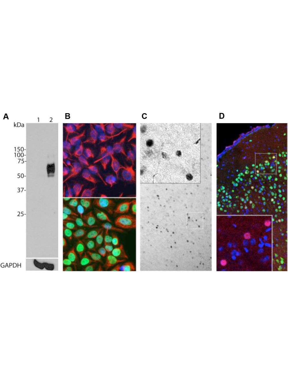
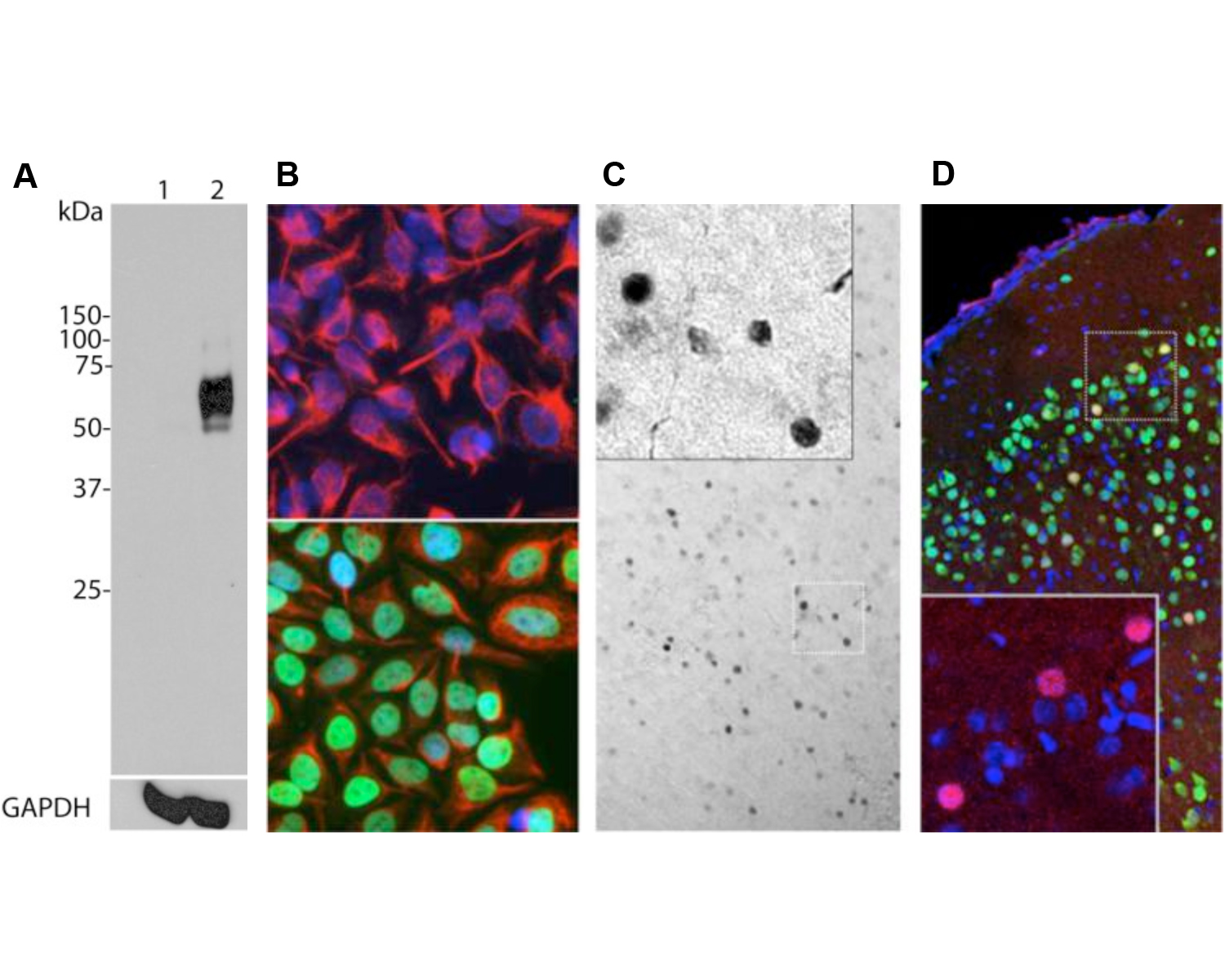
A: Western blot analysis of c-Fos expression in HeLa cells using M-1752-100. HeLa cells were serum-starved for 36 hours (Lane 1). Serum-starved HeLa cells were stimulated with 20% FBS for 2 hours (Lane 2). M-1752-100 recognizes bands in the range of 50-65 kDa, which represent multiple forms of c-Fos. A loading control was performed by stripping and re-probing the membrane with a monoclonal antibody against GAPDH, M-1376-250. B: Immunofluorescence staining of HeLa cells with M-1752-100. c-Fos staining (green) only localizes in the nuclei of 20% FBS stimulated cells (bottom panel), but not in un-stimulated cells (top panel). Cells were counter-stained with Chicken polyclonal antibody against vimentin, C-1409-50 (red) and DAPI (blue). C: Immunohistochemistry using M-1752-100 on 4% PFA transcardial-perfused mouse brain sections (45 uM thickness). c-FOS immunoreactive cells (dark colour, localized in cell nucleus) were visualized using a standard HRP-DAB (horseradish peroxidase-3,3'-diaminobenzidine) staining technique. D: Immunohistochemistry using M-1752-100 (red) and Rabbit polyclonal anti-NeuN/Fox3 (R-3770-100, green) on mouse cortical sections. Neurons positive for c-Fos and Fox3/NeuN appear yellow. The insert shows an enlarged image of staining with M-1752-100. Nuclei were labeled with DAPI (blue).
Western blot analysis of c-Fos expression in rat C6 cells (40 ug lysate loading per lane). C6 cells were serum-starved for 20 hours and then stimulated with 20% FBS for 2 hrs (+). Control cells were left in serum-depleted culture medium (-). M-1752-100 and R-1751-50 detect a strong band at 50 kDa in stimulated cells but not in control cells, representing increased c-FOS expression. Western Blotting Method: SDS-PAGE: denaturing and reducing, 4-12% Bis-Tris gel; Transfer: Tris-Glycine (Towbin's) buffer with 20% methanol; Membrane: PVDF (0.45 µm); Blocking: 5% skim milk in TBST, 1 hour at RT; Primary antibodies: R-1751-50 (1 µg/mL), M-1752-100 (1 µg/mL), incubation overnight at 4°C; Secondary antibodies: anti-rabbit-HRP (1/6000) or anti-mouse-HRP (1/3000), 1 hour at RT; Detection: Chemiluminiscence.
Left: Rat hippocampus stained for c-FOS (red) by Immunohistochemistry. Section was co-stained with rabbit antibody to FOX3/NeuN (R-3770-100, green). Blue: DAPI nuclear stain. The hippocampal neurons stain green for FOX3/NeuN and a few also are expressing c-FOS, and thus appear orange. These cells were spontaneously active at the time the animal was sacrificed. Right: Western blot analysis of c-FOS expression (green) in cell lysates. Mouse antibody to c-FOS was used at 1:1,000 dilution. GAPDH (red, lanes 2-5) was used as loading control (rabbit antibody to GAPDH, R-1701-100, 1:20,000). [1] protein standard, [2] HeLa cells in serum free media, [3] HeLa cells stimulated with 20% FBS for 2 hours after 36 hours serum starvation, [4] rat cortical neurons, [5] rat cortical neurons treated with membrane depolarization buffer for 5 hours. Multiple bands at 50-65 kDa in stimulated or treated cell lysates correspond to different forms of the c-Fos protein. The single band at 37 kDa (red) represents GAPDH protein.
Specific ReferencesChoi S et al. (2020). Parallel ascending spinal pathways for affective touch and pain. Nature. 587(7833):258-263. Application: IHC. Species: Mouse.
Bai L et al. (2019). Genetic Identification of Vagal Sensory Neurons That Control Feeding. Cell. 179(5):1129-43. Application: IHC. Species: Mouse.
General ReferencesVanstraaten et al (1983) Proc. Natl. Acad. Sci. 80: 3183 (Original molecular c-fos sequence paper)
Minson J. et al. (1994) Brain Res. 646: 44-52 (Early IHC localization paper)
 1800 605-5127
 +61 (0)8 8352 7711