Product DescriptiongoogleMouse anti-Spectrin alpha chain, non-erythrocytic 1 Monoclonal Antibody (Unconjugated), suitable for WB, IHC-Frozen, ICC, FC.
Alternative NamesSPTAN1; alpha II spectrin; alpha Fodrin; Spectrin, non-erythroid alpha chain; SPTA2;
Application(s)FC, ICC, IHC-Frozen, WB
Antibody HostMouse
Antibody TypeMonoclonal
SpecificityHuman; bovine; porcine; rat;
Species ReactivityBovine, Human, Mouse, Pig, Rat
Immunogen DescriptionThis antibody was raised against a recombinant construct containing the seventh, eight and ninth repeats (amino acids 676-1043) of human alpha-II Spectrin. The 9th spectrin repeat also includes a Src-homology 3 domain. This construct was expressed in and purified from E. coli.
Product DescriptionMouse anti-Spectrin alpha chain, non-erythrocytic 1 Monoclonal Antibody (Unconjugated), suitable for WB, IHC-Frozen, ICC, FC.
Application(s)FC, ICC, IHC-Frozen, WB
Application DetailsWB, ICC, IHC and FC. Recommended dilution of 1:1,000-1:2,000 for ICC and IHC, and 1:5,000-10,000 for WB. The protein is seen as a major band at 240 kDa depending on the species. For Flow Cytometry, use ~ 2 μg antibody per ~10^6 cells. Optimal concentrations/dilutions should be determined by the end-user.
TargetSpectrin alpha chain, non-erythrocytic 1
SpecificityHuman; bovine; porcine; rat;
Target Host SpeciesHuman
Species ReactivityBovine, Human, Mouse, Pig, Rat
Antibody HostMouse
Antibody TypeMonoclonal
Antibody IsotypeIgG1
Clone Name3D7
ConjugateUnconjugated
Immunogen DescriptionThis antibody was raised against a recombinant construct containing the seventh, eight and ninth repeats (amino acids 676-1043) of human alpha-II Spectrin. The 9th spectrin repeat also includes a Src-homology 3 domain. This construct was expressed in and purified from E. coli.
Purity DescriptionProtein G purified
FormatLyophilized from PBS buffer pH 7.2-7.6 with 0.1% trehalose, and sodium azide
Reconstitution InstructionsSpin vial briefly before opening. Reconstitute with 100 µL sterile-filtered, ultrapure water to achieve a 1 mg/mL concentration. Centrifuge to remove any insoluble material.
Storage InstructionsAliquot and store at -20°C for a higher stability and at 2-8°C with an appropriate antibacterial agent. Avoid freeze-thaw cycles.
Batch NumberPlease see item label.
Expiration Date12 months after date of receipt (unopened vial).
Alternative NamesSPTAN1; alpha II spectrin; alpha Fodrin; Spectrin, non-erythroid alpha chain; SPTA2;
Scientific BackgroundSpectrins are a family of filamentous cytoskeletal proteins that function as essential scaffold proteins that stabilize the plasma membrane and organize intracellular organelles. The Spectrins form into dimers and further into tetramers of alpha and beta subunits (Ref: Entrez Gene). The alpha-II subunit is widely expressed in tissues but, in the nervous system, is found predominantly in neurons.
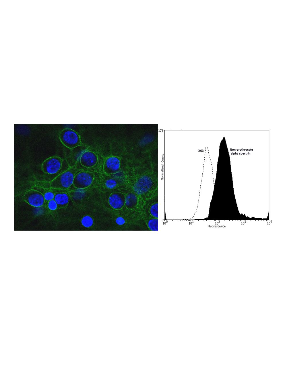
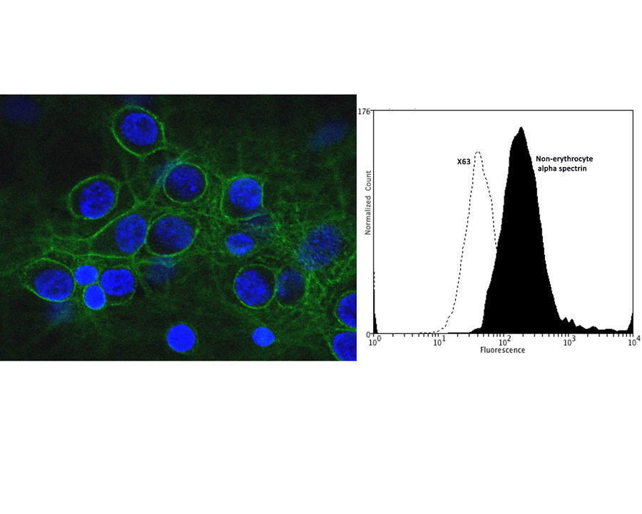
Left: Confocal image of mixed neuron-glial cultures stained with Mouse monoclonal antibody to non-erythrocyte alpha-spectrin M-1575-100 (green) and counterstained for DNA (blue). M-1575-100 stains numerous axonal and dendritic profiles in these cultures, and this image shows an optical section through a group of neuronal cell bodies. M-1575-100 clearly reveals the submembraneous cytoskeleton. Since alpha-II spectrin is specific for neurons in the CNS, the glial cells in this culture are not recognized by this antibody. This antibody also reveals the submembranous cytoskeleton of the axon. Right: Analysis of non-erythrocyte alpha spectrin expression in human neuroblastoma SH-SY5Y cell line by Flow Cytometry. Fixing and Permeabilization of cells: Absolute methanol (10 minutes in ice) and 0.1% Tween-20 in PBS, Blocking: 1% BSA, Primary antibody: Mouse Monoclonal antibody to Non-erythrocyte alpha spectrin (cat # M-1575-100, 2 μg per ~10^6 cells) for 30 minutes at room temperature, Secondary antibody: Goat anti-mouse PE labeled secondary antibody (1:100 fold dilution) with incubation for 20 minutes in dark at room temperature. Non-specific Control IgG, clone X63 (cat # M-1249-100) was used as negative control under same conditions (black dashed). Flow cytometry data and results were generated using Orflo MoxiflowTM instrument and protocols.
Left: Rat cerebellum stained for alpha II spectrin (green, 1:2,000) and GFAP (C-1373-50, red, 1:5,000). The spectrin antibody stains the submembraneous cytoskeleton on neurons and strongly reveals the cell bodies and dendrites of Purkinje cells, while the GFAP antibody stains the processes of Bergmann glia and astrocytes. Right: Western blot analysis of spectrin expression (green) in neural tissue and cell lysates. [1] protein standard, [2] rat whole brain, [3] rat spinal cord, [4] mouse whole brain, [5] mouse spinal cord, [6] NIH-3T3, [7] HEK293, [8] HeLa, [9] SH-SY5Y, [10] C6 glioma cells. A prominent band at about 250-260 kDa represents the intact alpha II spectrin heavy chain.
 1800 605-5127
 +61 (0)8 8352 7711