Product NameHigh-mobility group protein 1 (HMGB1), Mouse Monoclonal Antibody
Product DescriptiongoogleMouse anti-High-mobility group protein 1 (HMGP-1) Monoclonal Antibody (Unconjugated), suitable for WB, ICC, FC.
Application(s)FC, ICC, WB
Antibody HostMouse
Antibody TypeMonoclonal
SpecificityThe antibody reacts with a band at ~25 kDa by Western blot on HeLa cell extract. It has also been used successfully for immunocytochemistry showing strong nuclear staining.
Species ReactivityBovine, Human, Mouse, Other Mammals (Predicted), Pig, Rat
Immunogen DescriptionHuman full length recombinant human HMGB1 protein expressed in and purified from E. coli.
Product DescriptionMouse anti-High-mobility group protein 1 (HMGP-1) Monoclonal Antibody (Unconjugated), suitable for WB, ICC, FC.
Application(s)FC, ICC, WB
Application DetailsWestern Blotting (WB), Immunocytochemistry (ICC) and Flow Cytometry. A dilution of 1:1,000 - 1:2,000 is recommended for WB. A dilution of 1:500 - 1:1,000 is recommended for ICC. For Flow Cytometry, use ~2 ug per 10^6 cells. Biosensis recommends optimal dilutions/concentrations should be determined by the end user.
TargetHigh-mobility group protein 1 (HMGP-1)
SpecificityThe antibody reacts with a band at ~25 kDa by Western blot on HeLa cell extract. It has also been used successfully for immunocytochemistry showing strong nuclear staining.
Target Host SpeciesHuman
Species ReactivityBovine, Human, Mouse, Other Mammals (Predicted), Pig, Rat
Antibody HostMouse
Antibody TypeMonoclonal
Antibody IsotypeIgG2b
Clone Name1F3
ConjugateUnconjugated
Immunogen DescriptionHuman full length recombinant human HMGB1 protein expressed in and purified from E. coli.
Purity DescriptionProtein G purified
FormatLyophilized from PBS buffer pH 7.2-7.6 with 0.1% trehalose, and sodium azide
Reconstitution InstructionsSpin vial briefly before opening. Reconstitute with 100 µL sterile-filtered, ultrapure water to achieve a 1 mg/mL concentration. Centrifuge to remove any insoluble material.
Storage InstructionsAfter reconstitution of lyophilized antibody, aliquot and store at -20°C for a higher stability. Avoid freeze-thaw cycles.
Batch NumberPlease see item label.
Expiration Date12 months after date of receipt (unopened vial).
Scientific BackgroundHigh-mobility group proteins were named originally since they are abundand relatively low molecular weight proteins which run quickly on SDS-PAGE gels. High-mobility group protein box 1 (HMGB1, Amphoterin) is one of these. The "bx" in the name refers to the so-called high mobility group (HMG) box, a compact domain involved in DNA binding and protein-protein interactions. the HMGB1 molecule has two of these HMG domains. The protein is alslo called amphoterin, this name being derived from the presence of two highly charged regions in the molecule, a relatively neutrally charged N-terminus and a very negatively charged C-terminus. In fact the molecule is very unusually charged throughout, the human sequence consisting of 16.7% Glutamic acid, 9.3% Aspartic acid, 20% lysine and 9.3% Arginine. HMGB1 can bind Toll like receptor 4 (TLR4) and the Receptor for Advanced Glycation End products (RAGE). TLRs are components of the innate immune system, first recognized as a family of receptors which recognize "Pathogen Associated Molecular Pattern molecules (PAMPs). PAMPs are common components of bacteria and when TLRs bind these a strong inflammatory response is activated. More recently it has been recognized that TLRs can also be activated by Damage Associated Molecular Pattern molecules (DAMPs), which are endogenous substances released from damaged and diseased cells which also bind to TLR family receptors and also activate inflammation. HMGB1 is such a DAMP, binding to TLR4, and much evidence suggests that HMGB1 is a strong activator of inflammation. Interestingly, HMGB1 is released by necrotic cells but not by apoptotic cells (1).
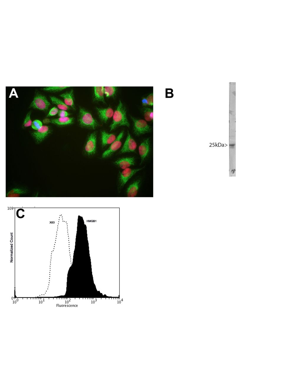
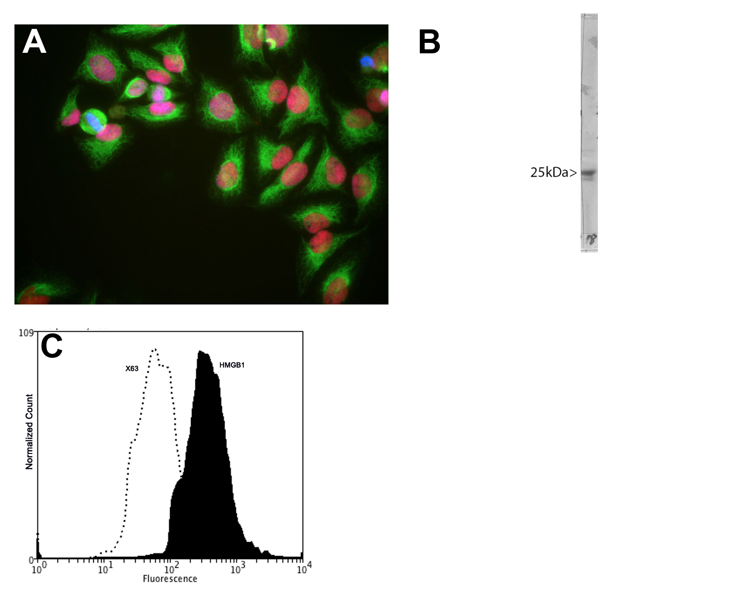
A: HeLa cells stained with M-1702-100 (red), chicken polyclonal antibody to Vimentin (C-1409-50, green) and DNA (blue). The M-1702-100 antibody reveals strong nuclear staining which overlaps with the DNA stain. B: Blot of crude HeLa cell extract stained with M-1702-100. HMGB1 runs at an apparent molecular weight of 25 kDa. C: Analysis of HMGB1 expression in human euroblastoma SH-SY5Y cell line by Flow Cytometry. Fixing and Permeabilization of cells: Absolute methanol (10 minutes in ice) and 0.1% Tween-20 in PBS, Blocking: 1% BSA, Primary antibody: Mouse Monoclonal antibody to HMGB1 (cat # M-1702-100, 2μg per ~10^6 cells) for 30 minutes at room temperature, Secondary antibody: Goat anti-mouse PE labeled secondary antibody (1:100 fold dilution) with incubation for 20 minutes in dark at room temperature. Non-specific Control IgG, clone X63 (cat # M-1249-200) was used as negative control under same conditions (black dashed). Flow cytometry data and results were generated using Orflo MoxiflowTM instrument and protocols.
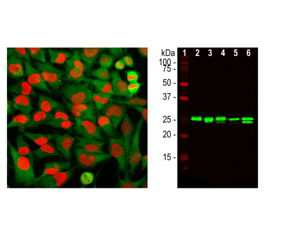
Left: HMGB1 detection in HeLa cells by Immunocytochemistry. Cells were stained with mouse antibody to HMGB1 (red, 1:2,000 ), and co-stained with rabbit antibody to GAPDH (R-1701-100, green, 1:2,000). The HMGB1 antibody stains the chromatin binding protein HMGB1, which is localized in the nuclei. In contrast, the GAPDH antibody produces strong cytoplasmic staining. Right: Western blot analysis of HMGB1 expression (green) in cell lysates, using mouse antibody to HMGB1 at 1:2,000 dilution. [1] protein standard, [2] NIH-3T3, [3] C6, [4] HEK293, [5] HeLa, and [6] SH-SY5Y. The 25 kDa band corresponds to HMGB1 protein.
General ReferencesScaffidi P, Misteli T, Bianchi ME. Release of chromatin protein HMGB1 by necrotic cells triggers inflammation. Nature 418:191-195 (2002).
 1800 605-5127
 +61 (0)8 8352 7711