Application DetailsThis antibody is recommended for WB, IHC, sandwich ELISA, immunofluorescence, Flow Cytometry. The recommended dilution for this antibody is 3 µg/mL for IHC and 10 µg/mL for immunofluorescence. Use ~2 ug per 10^6 cells for Flow Cytometry. Biosensis recommends optimal dilutions/concentrations should be determined by the end user.
SpecificitySpecificity has been confirmed by WB and direct ELISA against the antigen. Human. Other species have not been tested.
Target Host SpeciesHuman
Species ReactivityHuman
Antibody HostMouse
Antibody TypeMonoclonal
Antibody IsotypeIgG2a
Clone Name3H8
ConjugateUnconjugated
Immunogen DescriptionPartial recombinant human NeuroD1 (201-300) with a GST tag.
Purity DescriptionProtein G purified immunoglobulin
FormatLyophilized from PBS pH 7.2
Reconstitution InstructionsSpin vial briefly before opening. Reconstitute in 100 µL sterile-filtered, ultrapure water. Centrifuge to remove any insoluble material.
Storage InstructionsAfter reconstitution keep aliquots at -20°C for higher stability or at 2-8°C with an appropriate antibacterial agent. Glycerol (1:1) may be added for additional stability. Avoid repetitive freeze/thaw cycles.
Batch NumberPlease see item label.
Expiration Date12 months after date of receipt (unopened vial).
Alternative NamesNeurogenic differentiation factor 1; NeuroD; NEUROD1; NEUROD
NeuroD1 is a differentiation factor required for dendrite morphogenesis and maintenance in the cerebellar cortex. It is a transcriptional activator and binds to the insulin gene E-box. Efficient DNA binding requires dimerisation with another basic helix-loop-helix protein. In islet cells, NeuroD1 is phosphoryated on Ser-274 upon glucose stimulation and in activated neurons it is phosphorylated on Ser-335, which promotes dendritic growth. Defects in NeuroD1 cause maturity onset diabetes of the young type 6 (MODY6). It is characterised by the onset of diabetes during young adulthood and a primary defect in insulin secretion.
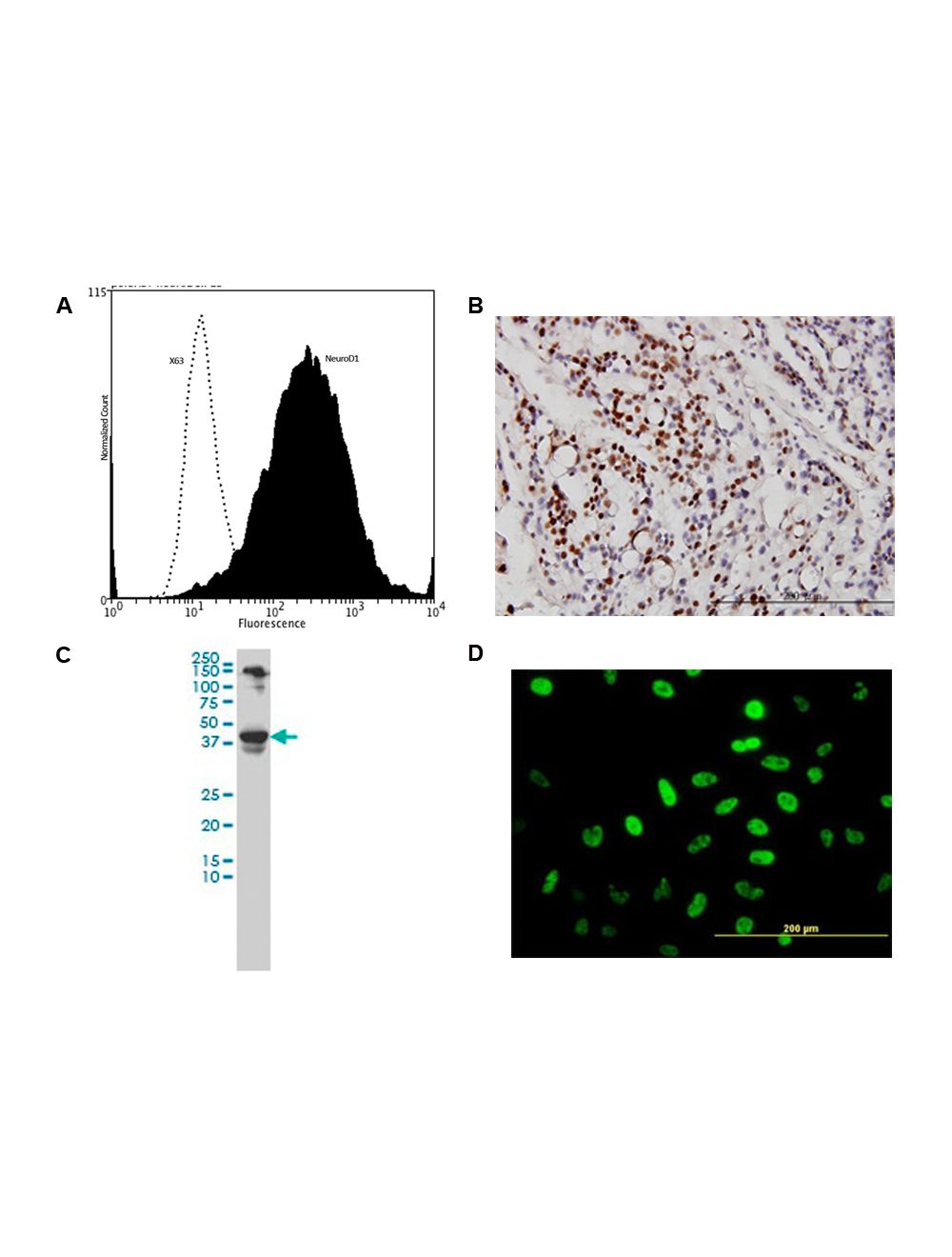
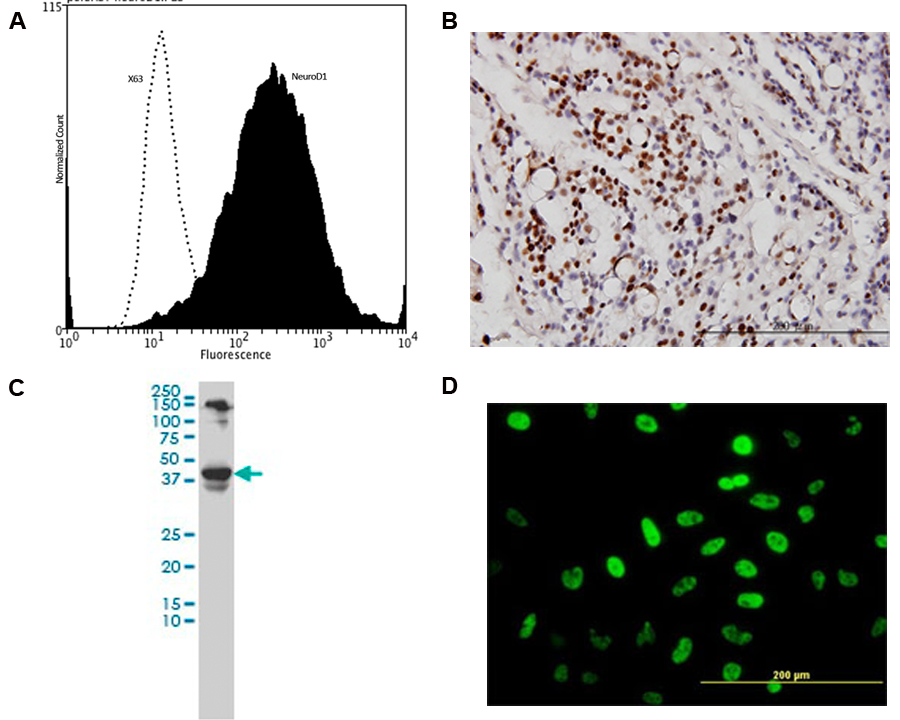
A: Specific staining of NeuroD1 expressed in human neuroblastoma SH-SY5Y cell line by Flow Cytometry using cat # M-850-100. Fixing and Permeabilization of cells: Absolute methanol (10 minutes in ice) and 0.1% Tween-20 in PBS, Blocking: 200 µg/mL Normal Sheep IgG (20 minutes), Primary antibody: Mouse Monoclonal antibody to NeuroD1 (2 ug per ~10^6 cells) for 30 minutes at room temperature, Secondary antibody: Goat anti-mouse PE labeled secondary antibody (1:100 dilution), 20 minutes in dark at room temperature. Negative control: Non-specific Control IgG, clone X63 (cat # M-1249-200, black dashed). Data and results were generated using Orflo MoxiflowTM instrument and protocols. B: Immunohistochemical detection of NeuroD1 on formalin fixed, paraffin-embedded, human ovary, clear cell carcinoma. Anti-NeuroD1 primary antibody was used at a concentration of 3 µg/mL. C: Western blot detection of NeuroD1 expression in human neuroblastoma cell lysate. D: Immunofluorescent detection of NeuroD1 in HeLa cells. Antibody concentration: 10 µg/mL.
 1800 605-5127
 +61 (0)8 8352 7711