1800 605-5127
 +61 (0)8 8352 7711
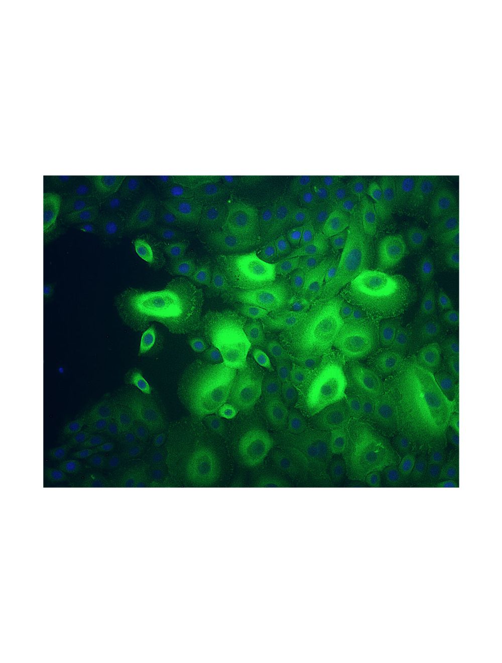

Vinculin, Mouse Monoclonal Antibody
As low as
US$427.00
Only %1 left
Catalog Number
M-1228
- Product Name Vinculin, Mouse Monoclonal Antibody
- Product Description Mouse anti-Vinculin Monoclonal Antibody (Unconjugated), suitable for WB, IHC-Frozen, IHC-Paraffin-embedded, ICC.
- Alternative Names Metavinculin; VCL;
- Application(s) ICC, IHC-Frozen, IHC-Paraffin-embedded, WB
- Antibody Host Mouse
- Antibody Type Monoclonal
- Specificity The specificity of this antibody has been confirmed by WB and IHC against the antigen. Human; mouse; rat; chicken;
- Species Reactivity Chicken, Human, Mouse, Rat
- Immunogen Description Purified Vinculin from human uterus
- Conjugate Unconjugated
- Purity Description IgG
- Regulatory Status For research use only.
Product Info
- Product Description Mouse anti-Vinculin Monoclonal Antibody (Unconjugated), suitable for WB, IHC-Frozen, IHC-Paraffin-embedded, ICC.
- Application(s) ICC, IHC-Frozen, IHC-Paraffin-embedded, WB
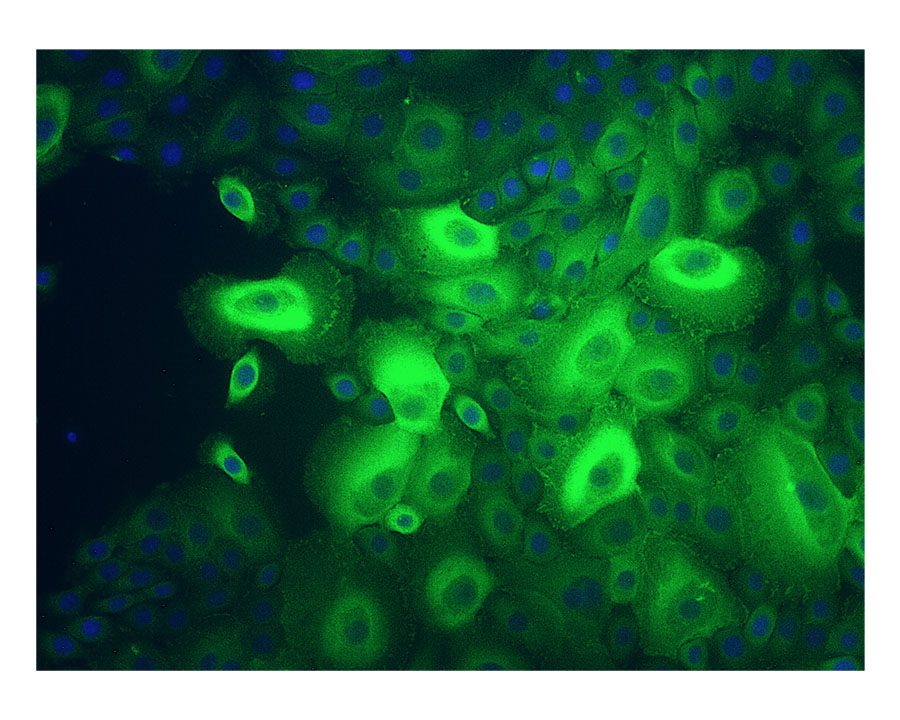
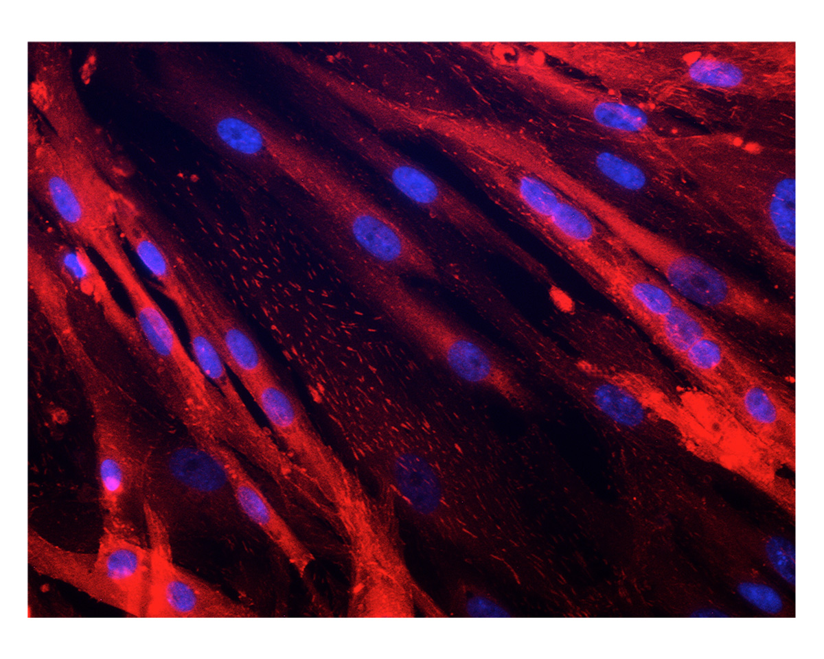
- Application Details Immunohistochemistry (IHC), Immunocytochemistry (ICC) and Western Blotting (WB). A concentration of 1.0-2.0 µg/mL is recommended for WB. Human Vinculin (isoform 2, Metavinculin) has a predicted length of 1,134 residues and a MW of 124 kDa. A concentration of 2.0-4.0 µg/mL is recommended to detect the protein in formalin/acetone fixed frozen tissues. A concentration of 2.0 µg/mL is recommended for IC. Biosensis recommends optimal dilutions/concentrations should be determined by the end user.
- Target Vinculin
- Specificity The specificity of this antibody has been confirmed by WB and IHC against the antigen. Human; mouse; rat; chicken;
- Target Host Species Human
- Species Reactivity Chicken, Human, Mouse, Rat
- Antibody Host Mouse
- Antibody Type Monoclonal
- Antibody Isotype IgG
- Clone Name VIN-54
- Conjugate Unconjugated
- Immunogen Description Purified Vinculin from human uterus
- Purity Description IgG
- Format Lyophilized from 1.2% sodium acetate, 2 mg BSA, 0.01 mg NaN3
- Reconstitution Instructions Spin vial briefly before opening. Reconstitute in 1 mL of sterile-filtered PBS (pH 7.4) to achieve an antibody concentration of 100 µg/mL. Centrifuge to remove any insoluble material.
- Storage Instructions At least 12 months after purchase at 2-8°C (lyophilized formulations). After reconstitution, aliquot and store at -20°C for a higher stability. Avoid freeze-thaw cycles.
- Batch Number Please see item label.
- Expiration Date 12 months after date of receipt (unopened vial).
- Alternative Names Metavinculin; VCL;
- Uniprot Number P18206
- Uniprot Number/Name P18206 (VINC_HUMAN)
- Scientific Background Vinculin is a cytoskeletal protein associated with cell-cell and cell-matrix adherens type junctions. Multiple isoforms are produced by alternative splicing.
- Shipping Temperature 25°C (ambient)
- UNSPSC CODE 41116161
- Regulatory Status For research use only.