Alternative NamesMyosin-7; Myosin heavy chain 7; Myosin heavy chain slow isoform; MyHC-slow; Myosin heavy chain, cardiac muscle beta isoform; MyHC-beta
SpecificitySlow myosin heavy chain. Clearly identifies Type 1 fibers. Within skeletal muscle clone is specific for slow myosin heavy chain in a wide variety of species. It reacts strongly with rat and feline slow myosin heavy chain. Clone also identifies beta (slow) myosin heavy chain in heart ventricles.
Species ReactivityCat, Chicken, Human, Mouse, Rabbit, Rat
Immunogen Description Human skeletal muscle myosin purified from myofibrils (Draeger A et al 1987;PMID: 2445924)
Application DetailsImmunohistochemistry (IHC frozen and paraffin-embedded) and Western Blotting (WB). A concentration of 0.5-2.0 µg/mL is recommended for WB. A concentration of 1.0-2.0 µg/mL is recommended to detect the protein in paraffin embedded sections by IHC. Heat-mediated antigen retrieval is required. Biosensis recommends optimal dilutions/concentrations should be determined by the end user.
TargetMyosin-7
SpecificitySlow myosin heavy chain. Clearly identifies Type 1 fibers. Within skeletal muscle clone is specific for slow myosin heavy chain in a wide variety of species. It reacts strongly with rat and feline slow myosin heavy chain. Clone also identifies beta (slow) myosin heavy chain in heart ventricles.
Target Host SpeciesHuman
Species ReactivityCat, Chicken, Human, Mouse, Rabbit, Rat
Antibody HostMouse
Antibody TypeMonoclonal
Antibody IsotypeIgG1
Clone NameNOQ7.5.4D
ConjugateUnconjugated
Immunogen Description Human skeletal muscle myosin purified from myofibrils (Draeger A et al 1987;PMID: 2445924)
Purity DescriptionRaw ascites
FormatLyophilized from mouse ascites fluid containing 1.2% sodium acetate, 2 mg BSA, and 0.01 mg NaN3 as preservative.
Reconstitution InstructionsSpin vial briefly before opening. Reconstitute in 100 uL sterile-filtered, ultrapure water. Centrifuge to remove any insoluble material.
Storage InstructionsStore lyophilized antibody at 2-8°C. After reconstitution keep aliquots at -20°C for a higher stability, or at 2-8°C for one month. Avoid repetitive freeze/thaw cycles.
Batch NumberPlease see item label.
Expiration Date12 months after date of receipt (unopened vial).
Alternative NamesMyosin-7; Myosin heavy chain 7; Myosin heavy chain slow isoform; MyHC-slow; Myosin heavy chain, cardiac muscle beta isoform; MyHC-beta
Scientific BackgroundMuscle myosin is a hexameric protein that consists of 2 heavy chain subunits (MHC), 2 alkali light chain subunits (MLC) and 2 regulatory light chain subunits (MLC-2).Clone identifies slow myosin heavy chain. Clearly identifies Type 1 fibers. Within skeletal muscle clone is specific for slow myosin heavy chain in a wide variety of species. It reacts strongly with rat and feline slow myosin heavy chain. Clone also identifies beta (slow) myosin heavy chain in heart ventricles.
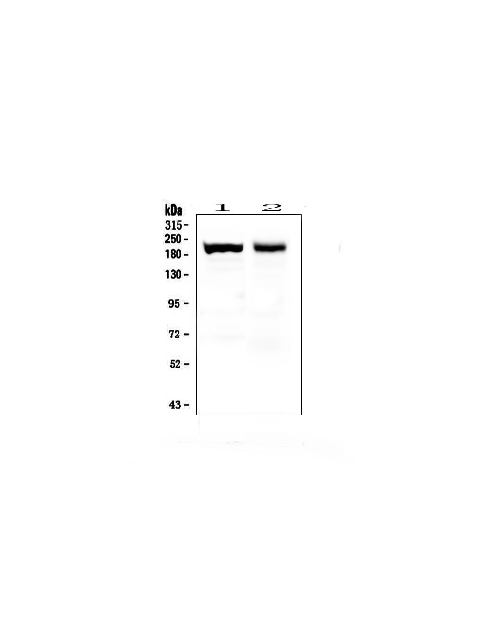
Western blot analysis of Myosin (Skeletal, Slow) using anti-Myosin (Skeletal, Slow) antibody (Catalog # M-1830-100). Electrophoresis was performed on a 5-20% SDS-PAGE gel at 70V (Stacking gel) / 90V (Resolving gel) for 2-3 hours. The sample well of each lane was loaded with 50ug of sample under reducing conditions. Lane 1: mouse skeletal muscle tissue lysates. Lane 2: rat skeletal muscle tissue lysates. After Electrophoresis, proteins were transferred to a Nitrocellulose membrane at 150mA for 50-90 minutes. The membrane was blocked with 5% Non-fat Milk/TBS for 1.5 hour at room temperature. The membrane was incubated with mouse anti- Myosin (Skeletal, Slow) antibody (Catalog # M-1830-100) at 0.5 µg/mL overnight at 4C, then washed with TBS-0.1% Tween20 3 times with 5 minutes each and probed with a goat anti-mouse IgG-HRP secondary antibody at a dilution of 1:10,000 for 1.5 hour at room temperature. A specific band was detected for Myosin (Skeletal, Slow) at approximately 200-220 kDa. The expected band size for Myosin (Skeletal, Slow) is at 220 kDa.
Slow skeletal myosin staining (red) in human myotubes by Immunocytochemistry. Primary antibody dilution: 1:500. Blue: Cell nuclei. Image courtesy of QBM Cell Science.
 1800 605-5127
 +61 (0)8 8352 7711