 1800 605-5127
 +61 (0)8 8352 7711
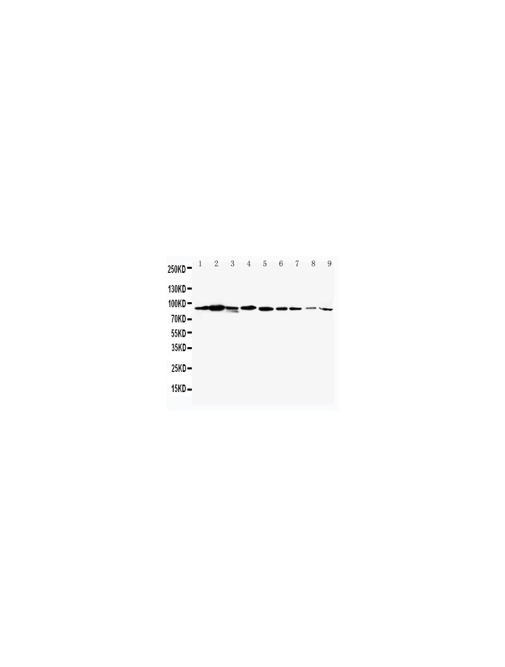

Signal transducer and activator of transcription 1-alpha (STAT1 alpha), Rabbit Polyclonal Antibody
- Product Name Signal transducer and activator of transcription 1-alpha (STAT1 alpha), Rabbit Polyclonal Antibody
-
Product Description
Rabbit anti-Signal transducer and activator of transcription 1-alpha (STAT1 alpha) Polyclonal Antibody (Unconjugated), suitable for WB.
-
Alternative Names
Transcription factor ISGF-3 components p91/p84; STAT1; p91.
- Application(s) WB
- Antibody Host Rabbit
- Antibody Type Polyclonal
-
Specificity
The specificity of this antibody has been confirmed by WB against the antigen. Human; mouse; rat; rabbit.
- Species Reactivity Human, Mouse, Rabbit, Rat
- Immunogen Description A synthetic peptide of human STAT1. To enhance the immunological response, this peptide was coupled to carrier protein BSA.
- Conjugate Unconjugated
- Purity Description Affinity purified on antigen column
- Regulatory Status For research use only.
Product Info
-
Product Description
Rabbit anti-Signal transducer and activator of transcription 1-alpha (STAT1 alpha) Polyclonal Antibody (Unconjugated), suitable for WB.
- Application(s) WB
- Application Details Western Blotting (WB). A concentration of 1.0-2.0 μg/mL is recommended for WB. Human STAT1 (isoform alpha/p91) has a predicted length of 750 residues and MW of 87 kDa. Biosensis recommends optimal dilutions/concentrations should be determined by the end user.
- Target Signal transducer and activator of transcription 1-alpha (STAT1 alpha)
-
Specificity
The specificity of this antibody has been confirmed by WB against the antigen. Human; mouse; rat; rabbit.
- Target Host Species Human
- Species Reactivity Human, Mouse, Rabbit, Rat
- Antibody Host Rabbit
- Antibody Type Polyclonal
- Antibody Isotype IgG
- Conjugate Unconjugated
- Immunogen Description A synthetic peptide of human STAT1. To enhance the immunological response, this peptide was coupled to carrier protein BSA.
- Purity Description Affinity purified on antigen column
- Format Lyophilized from a solution containing 5mg BSA, 0.9mg NaCl, 0.2mg Na2HPO4, 0.05mg Thimerosal, 0.05mg NaN3.
- Reconstitution Instructions Spin vial briefly before opening. Reconstitute with 200 μL sterile-filtered, ultrapure water to achieve a 0.5 mg/mL concentration. Centrifuge to remove any insoluble material.
- Storage Instructions After reconstitution of lyophilized antibody, aliquot and store at -20°C for a higher stability (up to 6 months). Avoid freeze-thaw cycles. Reconstituted antibody can be stored for 1 month at 2-8°C. Unopened, lyophilized vial may be stored for 12 months from date of receipt at -20°C.
- Batch Number Please see item label.
- Expiration Date 12 months after date of receipt (unopened vial).
-
Alternative Names
Transcription factor ISGF-3 components p91/p84; STAT1; p91.
- Uniprot Number P42224
- Uniprot Number/Name P42224 (STAT1_HUMAN)
- Scientific Background STAT1 belongs to the STAT (signal transducers and activators of transcription) family of proteins. STAT proteins undergo tyrosine/serine phosphorylation in response to cytokines and growth factors. As a result of this phosphorylation, STAT proteins form dimers and are translocated to the nucleus where they act as transcription factors. Alternative splicing of the STAT1 gene gives rise to the STAT1 alpha and beta isoforms.
- Shipping Temperature 2-8°C (on cold packs)
- UNSPSC CODE 41116161
- Regulatory Status For research use only.
