Alternative NamesBrain-derived neurotrophic factor; Abrineurin
Application(s)ELISA
Antibody HostRabbit
Antibody TypePolyclonal
SpecificityHuman, rat and mouse BDNF. Expected to detect BDNF from other species due to sequence homology. No cross-reactivity with other neurotrophins.
Species ReactivityHuman, Other Mammals (Predicted)
Immunogen DescriptionAntibody was raised against a GST-tagged rhBDNF fusion protein and expressed in and purified from E. coli.
ConjugateUnconjugated
Purity DescriptionAffinity purified on antigen column.
Application DetailsWestern Blotting (denaturing and reducing): 0.2 to 1 µg/mL. Antibody detects 14 kDa mature BDNF monomer and 32 kDa proBDNF monomer in cell lysate and tissue homonenate. Antibody has only been tested on cell lysate and tissue homogenate of human origin. Acid-treated samples may give cleaner blots, and enhance signals for BDNF. R-1707-100 is not recommended for human serum samples. For human serum analysis, we recommend mouse monoclonal antibody to rhBDNF (M-1744-50/100), or rabbit polyclonal antibody to BDNF peptide 1-10 (R-083-100, whole serum; R-066-500, IgG).
Flow Cytometry: ~2 µg per 10^6 cells, methanol fixation. Note: R-1707-100 cannot be used to distinguish the flow cytometry signal originating from mature BDNF versus proBDNF.
Biosensis recommends optimal dilutions/concentrations should be determined by the end user.
TargetBrain-derived neurotrophic factor (BDNF)
SpecificityHuman, rat and mouse BDNF. Expected to detect BDNF from other species due to sequence homology. No cross-reactivity with other neurotrophins.
Target Host SpeciesHuman
Species ReactivityHuman, Other Mammals (Predicted)
Antibody HostRabbit
Antibody TypePolyclonal
Antibody IsotypeIgG
ConjugateUnconjugated
Immunogen DescriptionAntibody was raised against a GST-tagged rhBDNF fusion protein and expressed in and purified from E. coli.
Purity DescriptionAffinity purified on antigen column.
FormatLyophilized from a solution containing PBS pH 7.4, 3% trehalose, with 0.05% sodium azide.
Reconstitution InstructionsSpin vial briefly before opening. Reconstitute vial with 100 µL sterile-filtered, ultrapure water to obtain a concentration of 1 mg/mL. Centrifuge to remove any insoluble material.
Storage InstructionsStore lyophilized antibody at 2-8°C protected from moisture. After reconstitution divide antibody into useful aliquots and keep aliquots at -20°C to -80°C for a higher stability. Working aliquots can be kept at 2-8°C for up to 1 month. Avoid repetitive freeze/thaw cycles.
Batch NumberPlease see item label.
Expiration Date12 months after date of receipt (unopened vial).
Alternative NamesBrain-derived neurotrophic factor; Abrineurin
Scientific BackgroundBDNF belongs to the neurotrophin family and promotes the survival of neuronal populations that are all located either in the central nervous system or directly connected to it. It is a major regulator of synaptic transmission and plasticity at adult synapses in many regions of the CNS. The versatility of BDNF is emphasized by its contribution to a range of adaptive neuronal responses including long-term potentiation (LTP), long-term depression (LTD), certain forms of short-term synaptic plasticity, as well as homeostatic regulation of intrinsic neuronal excitability. The alterations in BDNF expression induced by various kinds of brain insult including stress, ischemia, seizure activity and hypoglycemia, may contribute to some pathologies such as depression, epilepsy, Alzheimer's, and Parkinson's disease. Microglia release BDNF that may contribute to neuroinflammation and neuropathic pain. SUBUNIT: Monomers and homodimers. Binds to NTRK2/TRKB. SUBCELLULAR LOCATION: Secreted protein. POst translation modification: Converted into mature BDNF by plasmin (PLG). SIMILARITY: Belongs to the NGF-beta family.
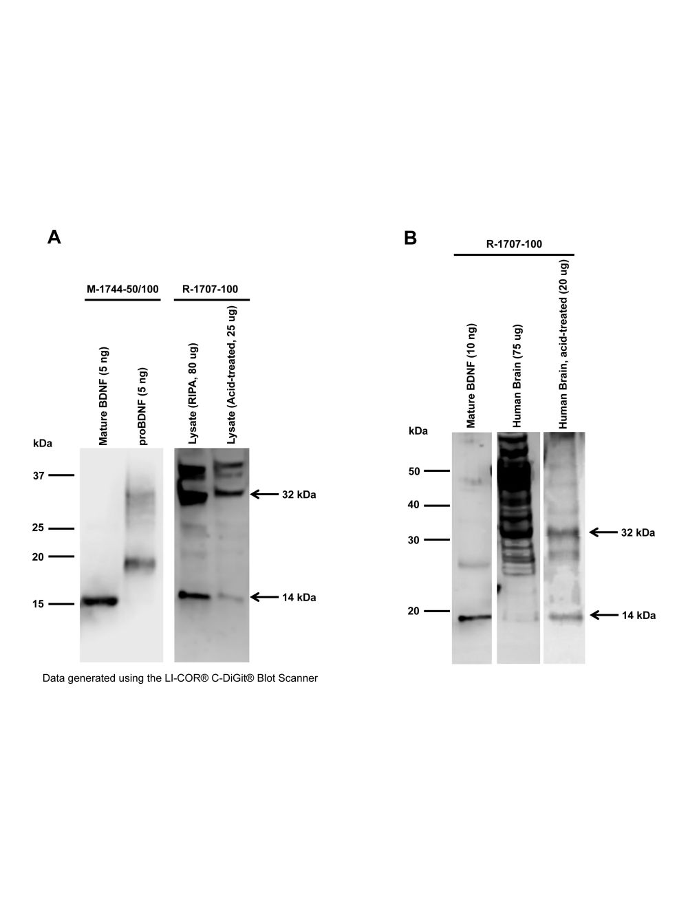
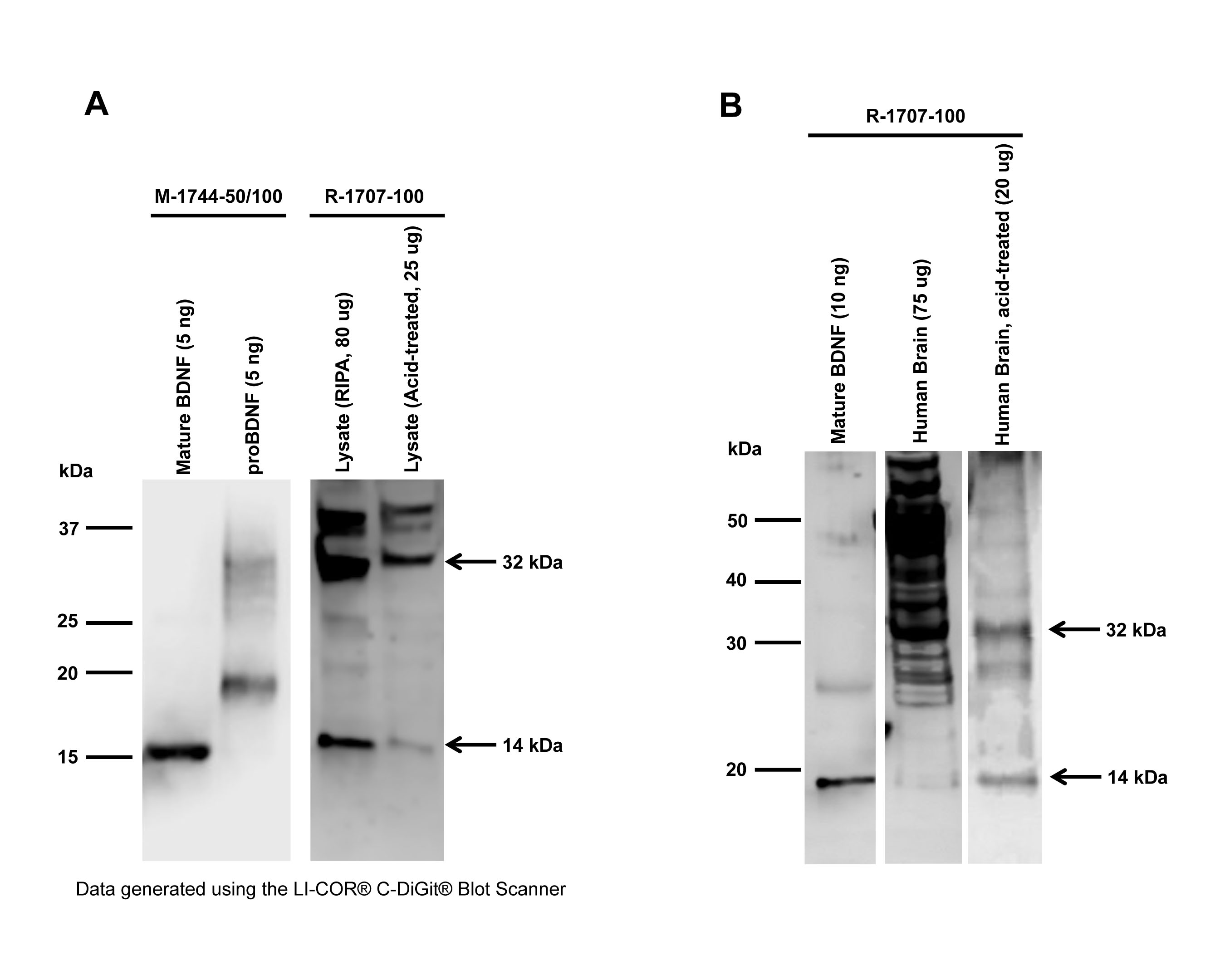
Western blot analysis of BDNF expression in human SH-SY5Y cell lysates (A) and human brain (B). Polyclonal rhBDNF antibody R-1707-100 (1 µg/mL) detects monomeric BDNF at 14 kDa, and monomeric proBDNF at 32 kDa in lysates prepared either in RIPA buffer or in acid-extraction buffer (A). Control antibody M-1744-50/100 (1 µg/mL) confirms detection of mature BDNF (Lane 1) and proBDNF (Lane 2) using BDNF and proBDNF proteins. A second band at 18 kDa is observed in the proBDNF standard, likely to represent a proBDNF degradation product. In human brain (B), R-1707-100 detects mature BDNF (14 kDa) and proBDNF (32 kDa) in both preparations (Tris-buffer and acid-treated). Acid-treated brain demonstrates lower background staining and gives a stronger BDNF signal.
Western Blotting Method: SDS-PAGE: denaturing and reducing, 12% Bis-Tris gel; Transfer: Tris-Glycine buffer, semi-dry transfer; Membrane: nitrocellulose (0.22 µm); Blocking: 5% skim milk in TBST, 1 hour at RT; Primary antibody: overnight at 4°C; Secondary antibody: anti-mouse-HRP or anti-rabbit-HRP (1/6000), 2 hours at RT; Detection: Chemiluminiscence.
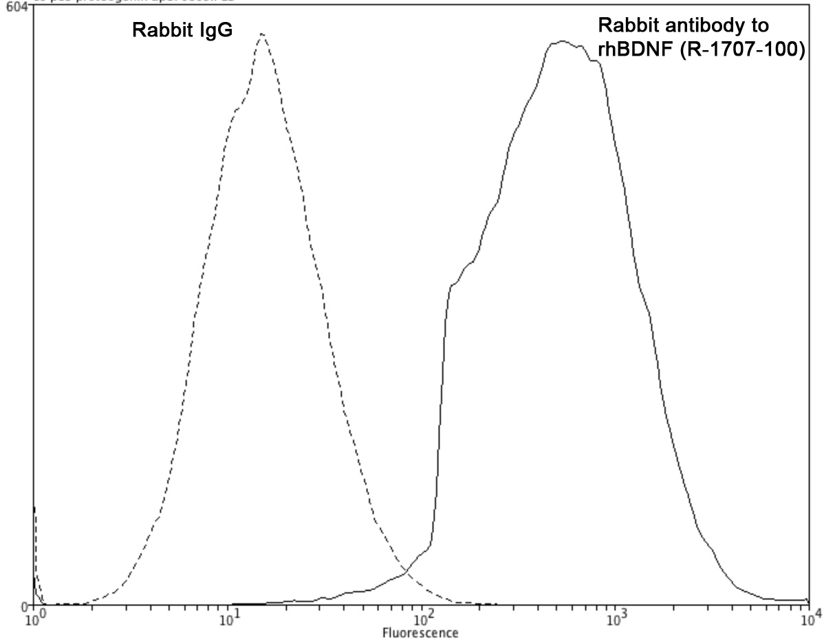
Analysis of BDNF expression in rat C6 cells by Flow Cytometry. Fixing and permeabilization of cells: Absolute methanol (10 minutes in ice) and 0.1% Tween-20 in PBS, Blocking: 200 µg/mL sheep IgG, Primary antibody: Rabbit polyclonal antibody to rhBDNF (cat # R-1707-100, 2 µg per ~10^6 cells) for 30 minutes at room temperature, Secondary antibody: Goat anti-rabbit-PE labeled secondary antibody (1:100 dilution), 20 minutes in dark at room temperature. Negative control: Normal rabbit IgG. Data and results were generated using Orflo MoxiflowTM instrument and protocols.
 1800 605-5127
 +61 (0)8 8352 7711