 1800 605-5127
 +61 (0)8 8352 7711
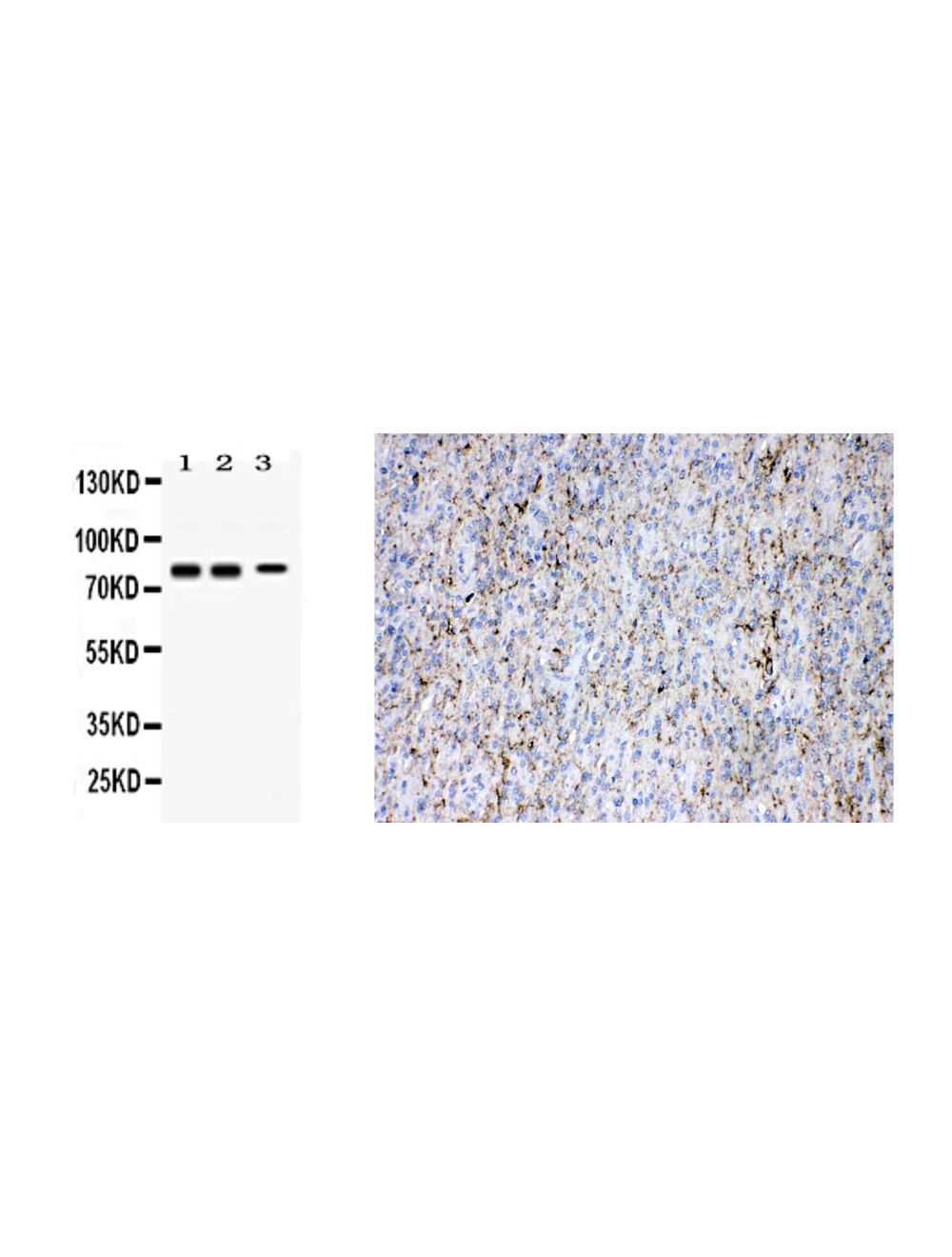

Synapsin-1 (SYN1), Rabbit Polyclonal Antibody
Only %1 left
Catalog Number
R-1829
Discontinued
- Product Name Synapsin-1 (SYN1), Rabbit Polyclonal Antibody
- Product Description Rabbit anti-Synapsin-1 (SYN1) Polyclonal Antibody (Unconjugated), suitable for WB, IHC-Paraffin-embedded.
- Alternative Names Synapsin-1; Brain protein 4.1; Synapsin I; SYN1
- Application(s) IHC-Paraffin-embedded, WB
- Antibody Host Rabbit
- Antibody Type Polyclonal
- Specificity This antibody is specific for Synapsin I as demonstrated by western blotting.
- Species Reactivity Human, Mouse, Rat
- Immunogen Description A synthetic peptide corresponding to a sequence at the C-terminus of human Synapsin I (662-705aa KSQSLTNAFNLPEPAPPRPSLSQDEVKAETIRSLRKSFASL FSD), identical to the related mouse and rat sequences.
- Conjugate Unconjugated
- Purity Description Affinity purified
- Regulatory Status For research use only.
Product Info
- Product Description Rabbit anti-Synapsin-1 (SYN1) Polyclonal Antibody (Unconjugated), suitable for WB, IHC-Paraffin-embedded.
- Application(s) IHC-Paraffin-embedded, WB
- Application Details Western Blot (0.1-0.5 µg/mL): tested in SHG-44 cell lysate (human malignant glioma cell line), mouse and rat brain lysates, and rat cell line. Immunohistochemistry (0.5-1.0 µg/mL): tested in formalin fixed, paraffin embedded human glioma tissue with HEIR. Other applications not yet tested. Biosensis recommends optimal dilutions/concentrations should be determined by the end user.
- Target Synapsin-1 (SYN1)
- Specificity This antibody is specific for Synapsin I as demonstrated by western blotting.
- Target Host Species Human
- Species Reactivity Human, Mouse, Rat
- Antibody Host Rabbit
- Antibody Type Polyclonal
- Antibody Isotype IgG
- Conjugate Unconjugated
- Immunogen Description A synthetic peptide corresponding to a sequence at the C-terminus of human Synapsin I (662-705aa KSQSLTNAFNLPEPAPPRPSLSQDEVKAETIRSLRKSFASL FSD), identical to the related mouse and rat sequences.
- Sequence KSQSLTNAFNLPEPAPPRPSLSQDEVKAETIRSLRKSFASLFSD
- Purity Description Affinity purified
- Format Lyophilized
- Reconstitution Instructions Spin vial briefly before opening. Reconstitute in 100 uL sterile-filtered, ultrapure water to prepare 1 mg/mL solution. Centrifuge to remove any insoluble material.
- Storage Instructions Store lyophilized antibody at 2-8°C. After reconstitution divide into aliquots and store at -20°C for long-term storage. Store at 2-8°C short-term (up to 4 weeks) with an appropriate antibacterial agent. Avoid repetitive freeze/thaw cycles.
- Batch Number Please see item label.
- Expiration Date 12 months after date of receipt (unopened vial).
- Alternative Names Synapsin-1; Brain protein 4.1; Synapsin I; SYN1
- Uniprot Number P17600
- Uniprot Number/Name P17600 (SYN1_HUMAN)
- Scientific Background The synapsins are a family of proteins that have long been implicated in the regulation of neurotransmitter release at synapses. Specifically, they are thought to be involved in regulating the number of synaptic vesicles available for release via exocytosis at any one time. Synapsins are present in invertebrates and vertebrates and are somewhat homologous across evaluated vertebrates. (Ref: Pfam.org)
- Shipping Temperature 25°C (ambient)
- UNSPSC CODE 41116161
- Regulatory Status For research use only.
