Product NameFluoro-Jade C (FJC) Powder for identifying Degenerating Neurons
Product DescriptiongoogleThe causes and effects of neuronal degeneration are of major interest to a wide variety of neuroscientists. Paralleling this growing interest is an increasing number of methods applicable to the detection of neuronal degeneration. Fluoro-Jade C stains all degenerating neurons regardless of specific insult or mechanism of cell death. Fluoro-Jade C exhibits the greatest signal to background ratio, as well as the highest resolution. This translates to a stain of maximal contrast and affinity for degenerating neurons. This makes it ideal for localising not only degenerating nerve cell bodies but also distal dendrites, axons and terminals. The dye is highly resistant to fading and is compatible with virtually all histological processing and staining protocols.
Note: This product is equivalent to discontinued product AG325 from Merck-Millipore.
Alternative NamesFJC, Fluoro-Jade
Application(s)FC, ICC
SpecificityDegenerating neurons, and neuronal degeneration. There is no specific staining in normal healthy brain. Note: Some researchers under some conditions report blood vessel staining with Fluoro Jade. This may be because Fluoro Jade is an analogue of eosin (which stains blood cells). In general, good perfusion and preparation of the tissue should help prevent blood vessel staining but it may not be possible to eliminate it entirely. In our experience it is generally possible to distinguish neuronal from blood vessels staining by eye.
Species ReactivityHuman, Mouse, Other Mammals (Predicted), Rat
Purity DescriptionSilica TLC (acetanitrile/water, 6/4) revealed the presence of 2 fluorescent spots, presumably corresponding to the mono- and di-sulphate homologues. The presence of precursors or free fluorescein was not detected.
Product DescriptionThe causes and effects of neuronal degeneration are of major interest to a wide variety of neuroscientists. Paralleling this growing interest is an increasing number of methods applicable to the detection of neuronal degeneration. Fluoro-Jade C stains all degenerating neurons regardless of specific insult or mechanism of cell death. Fluoro-Jade C exhibits the greatest signal to background ratio, as well as the highest resolution. This translates to a stain of maximal contrast and affinity for degenerating neurons. This makes it ideal for localising not only degenerating nerve cell bodies but also distal dendrites, axons and terminals. The dye is highly resistant to fading and is compatible with virtually all histological processing and staining protocols.
Note: This product is equivalent to discontinued product AG325 from Merck-Millipore.
Application DetailsFollowing our detailed protocol, Fluoro-Jade B labels degenerating neurons which are visualised with blue light excitation, while DAPI (not included) counter stains cell nuclei, visualised with ultra-violet illumination. The Fluoro-Jade C dye can be used on all kinds of preserved tissues, including fresh-frozen, paraformaldehyde or formalin fixed, and formalin fixed, paraffin-embedded tissues.
TargetDegenerating neurons
SpecificityDegenerating neurons, and neuronal degeneration. There is no specific staining in normal healthy brain. Note: Some researchers under some conditions report blood vessel staining with Fluoro Jade. This may be because Fluoro Jade is an analogue of eosin (which stains blood cells). In general, good perfusion and preparation of the tissue should help prevent blood vessel staining but it may not be possible to eliminate it entirely. In our experience it is generally possible to distinguish neuronal from blood vessels staining by eye.
Target Host SpeciesSpecies Independent
Species ReactivityHuman, Mouse, Other Mammals (Predicted), Rat
Ex/Em MaxFJC visualization is accomplished using blue light or a 488 nm Laser. Excitation Peak: 495 nm Emission Peak: 521 nm Filter system for visualizing: Fluorescein/FITC
Detection MethodFluorescence
Kit ComponentsMaterials Provided:
30 mg Fluoro-Jade C, dry powder Detailed protocol
Equipment and Reagents Required:
Distilled water ACS grade Ethanol (200 proof) for slide & solution preparation 1% sodium hydroxide in 80% ethanol (basic alcohol solution) 0.1% Acetic Acid solution (in water) 70% ethanol in distilled water 0.06% (KMnO4) potassium permanganate solution DAPI powder or 100X solution (working range is 0.5-5 µg/mL) Xylene liquid Staining dishes/Coplin jars Cover slips DPX mounting media or another permanent mounting medium. Non-polar media are preferred over aqueous mounting media such as glycerin/water to obtain high- contrast images (refer to Appendix B in the protocol for a comparative analysis). Traditional fluorescent mounting mediums are not recommended because of their high pH. Slide warmer Convection oven
Purity DescriptionSilica TLC (acetanitrile/water, 6/4) revealed the presence of 2 fluorescent spots, presumably corresponding to the mono- and di-sulphate homologues. The presence of precursors or free fluorescein was not detected.
FormatDry, Coffee brown to brick red powder; hygroscopic powder keep dessicated.
Reconstitution InstructionsSpin vial briefly before opening. See product protocol for detailed use instructions.
Storage InstructionsThe powdered dye can be stored desiccated at room temperature in the dark. Storage in a desiccator is recommended as FJB is hydroscopic. The 0.01% stock solution will remain stable for 3 months when stored in a refrigerator, in the dark. The 0.0001-0.0004% working solution in 0.1% acetic acid should be used within 4 hours of preparation. Diluted FJB dye solutions are not stable and should not be stored. The other diluted solutions can be reused and stored for up to 48 hours if refrigerated and protected from light. Best results require freshly diluted solutions.
Batch NumberPlease see item label.
Expiration Date6 months after date of receipt (unopened vial).
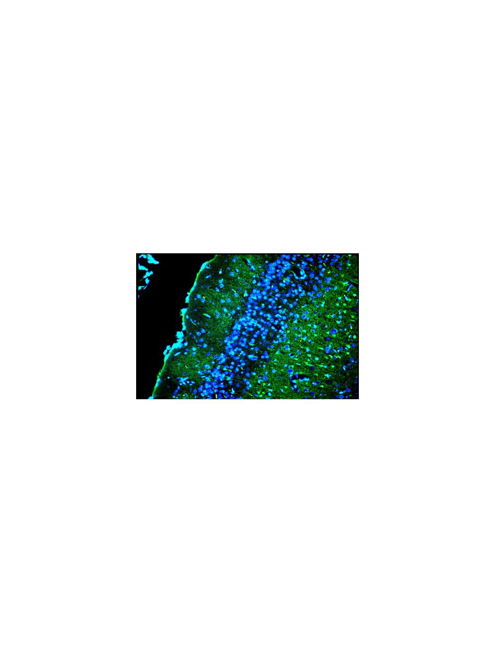
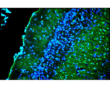
Double exposure using combined blue and ultraviolet epi-fluorescent illumination of the superficial layers of the cingulated rat cortex exposed to kainic acid. Layer I contains conspicuous Fluoro-Jade C positive degenerating axon terminals. Layer II contains densely packed DAPI-positive viable granule cells. Layer III contains a mixture of Fluoro-Jade C positive denegerating pyramidal cells and DAPI-positive viable pyramidal cells. Photo is courtesy of Dr. Larry Schmued.
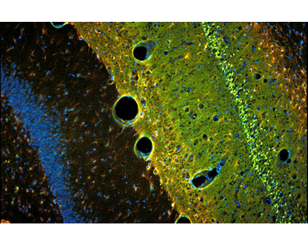
Triple exposure combining ultraviolet, blue and green light epi-fluorescent illumination (10X) of rat hippocampus exposed to kainic acid. The section was triple labeled with Fluoro-Jade C and DAPI staining combined with GFAP immunohistochemistry. The section reveals extensive green Fluoro-Jade C positive neuronal degeneration throughout the entire CA-1 region of the hippocampus. The underlying blue viable positive granule cells of the dentate gyrus are only DAPI positive. Both regions exhibit red GFAP positive hypertrophied astrocytes. Photo is courtesy of Dr. Larry Schmued.
 1800 605-5127
 +61 (0)8 8352 7711