Product DescriptiongoogleHQ-O Ready-To-Dilute (RTDTM) Stain Reagent is designed to label amyloid plaques in paraffin-embedded or freshly cut frozen tissue sections. As a fluorescent zinc chelator, HQ-O is unique as it takes advantage of the known presence of concentrated zinc in amyloid plaques. Studies with HQ-O revealed that fluorescent plaque-like structures are only seen when synthetic Aβx-42 is aggregated in the presence of zinc. Under blue light excitation, plaque structures appear bright green fluorescent in the brain parenchyma, correlating closely with plaque structures observed following Aβ antibody staining. HQ-O RTDTM staining reagent is compatible with other fluorophores, such as DAPI, Hoechst and ethidium bromide, as well as fluorescent-labelled antibodies with emission spectra in the blue and/or red emission range of fluorescent microscopes. Due to its zinc-chelating characteristics, HQ-O RTDTM staining reagent may visualize globular structures within blood vessels and intravascular leucocytes. HQ-O RTDTM staining reagent has multiple advantages over older blue-light exciting stains such as Thioflavin S. Thioflavin S typically exhibits relatively low contrast and resolution and suffers from bleed-through when excited by wavelengths other than blue light. HQ-O RTDTM staining reagent suffers none of these setbacks and not only provides a higher contrast and longer lasting dye, but because it lacks excitation bleed-through, HQ-O can be readily adapted to multiple labelling studies very easily. To visualize the HQ-O tracer, it is recommended to use a filter cube designed for visualizing Fluorescein/FITC or a blue-light laser. Although it can be seen with both narrow and wide-band pass filters, there is no need to use a narrow band filter since the compound does not bleed through when excited with other filters. A recommended excitation range of a wide band filter is 447-503 nm, with a peak at 475.
Alternative NamesHQ-O tracer
Application(s)IHC-Frozen, IHC-Paraffin-embedded
SpecificityAs a fluorescent zinc chelator, HQ-O is unique as it takes advantage of the known presence of concentrated zinc in amyloid plaques. Studies with HQ-O revealed that fluorescent plaque-like structures are only seen when synthetic Aβx-42 is aggregated in the presence of zinc. Under blue light excitation, plaque structures appear bright green fluorescent in the brain parenchyma, correlating closely with plaque structures observed following Aβ antibody staining.
Species ReactivityHuman, Mouse, Other Mammals (Predicted), Rat
Product DescriptionHQ-O Ready-To-Dilute (RTDTM) Stain Reagent is designed to label amyloid plaques in paraffin-embedded or freshly cut frozen tissue sections. As a fluorescent zinc chelator, HQ-O is unique as it takes advantage of the known presence of concentrated zinc in amyloid plaques. Studies with HQ-O revealed that fluorescent plaque-like structures are only seen when synthetic Aβx-42 is aggregated in the presence of zinc. Under blue light excitation, plaque structures appear bright green fluorescent in the brain parenchyma, correlating closely with plaque structures observed following Aβ antibody staining. HQ-O RTDTM staining reagent is compatible with other fluorophores, such as DAPI, Hoechst and ethidium bromide, as well as fluorescent-labelled antibodies with emission spectra in the blue and/or red emission range of fluorescent microscopes. Due to its zinc-chelating characteristics, HQ-O RTDTM staining reagent may visualize globular structures within blood vessels and intravascular leucocytes. HQ-O RTDTM staining reagent has multiple advantages over older blue-light exciting stains such as Thioflavin S. Thioflavin S typically exhibits relatively low contrast and resolution and suffers from bleed-through when excited by wavelengths other than blue light. HQ-O RTDTM staining reagent suffers none of these setbacks and not only provides a higher contrast and longer lasting dye, but because it lacks excitation bleed-through, HQ-O can be readily adapted to multiple labelling studies very easily. To visualize the HQ-O tracer, it is recommended to use a filter cube designed for visualizing Fluorescein/FITC or a blue-light laser. Although it can be seen with both narrow and wide-band pass filters, there is no need to use a narrow band filter since the compound does not bleed through when excited with other filters. A recommended excitation range of a wide band filter is 447-503 nm, with a peak at 475.
Application DetailsDetection and fluorescent-staining of amyloid plaques in paraffin-embedded or freshly cut frozen tissue sections, please see detailed protocol for specific use instructions.
TargetAmyloid plaque
SpecificityAs a fluorescent zinc chelator, HQ-O is unique as it takes advantage of the known presence of concentrated zinc in amyloid plaques. Studies with HQ-O revealed that fluorescent plaque-like structures are only seen when synthetic Aβx-42 is aggregated in the presence of zinc. Under blue light excitation, plaque structures appear bright green fluorescent in the brain parenchyma, correlating closely with plaque structures observed following Aβ antibody staining.
Target Host SpeciesSpecies Independent
Species ReactivityHuman, Mouse, Other Mammals (Predicted), Rat
Ex/Em MaxTo visualize the HQ-O tracer, it is recommended to use a filter cube designed for visualizing Fluorescein/FITC or a blue-light laser. Although it can be seen with both narrow and wide-band pass filters, there is no need to use a narrow band filter since the compound does not “bleed through” when excited with other filters. A recommended excitation range of a wide band filter is 447 – 503 nm, with a peak at 475 nm.
Detection MethodFluorescence
Kit ComponentsOne bottle containing 20 mL (TR-700-HQOT) or 40 mL (TR-700-HQO) of 10X HQ-O RTDTM solution This quantity will be sufficient for approximately:
TR-700-HQOT: 4 Coplin Jars or 1-2 staining dishes
TR-700-HQO: 8 Coplin Jars or 2-4 staining dishes
Storage InstructionsStore 10X stock solution at 2-8°C protected from light, for up to 6 months. The diluted dye (1X) should be used within 24 hours.
Batch NumberPlease see item label.
Expiration Date6 months after date of receipt (10X stock solution)
Alternative NamesHQ-O tracer
Scientific BackgroundA novel zinc chelator, HQ-O, was developed for localizing zinc within amyloid plaques. The histology involves incubating tissue sections in a dilute aqueous solution of HQ-O.
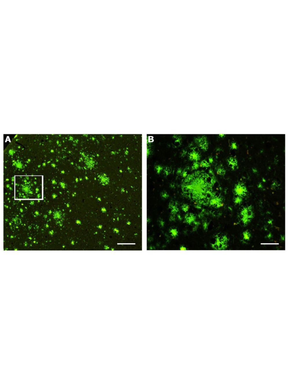
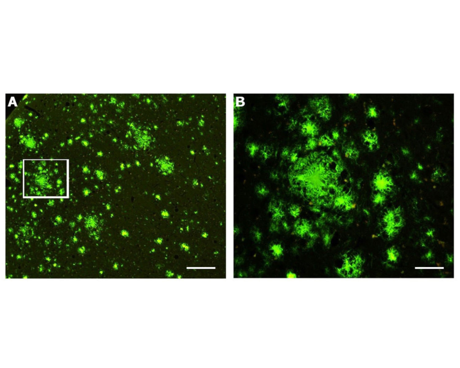
Labelling of amyloid plaques (green) in paraffin-processed cortex from a 1 year old APP/PSI mouse model of AD (A). B: High-magnification view of the cortical region outlined in white in A.
D: Within the CA4 region of the hippocampus, co-labeling using GFAP antibody demonstrates the relationship between hypertrophied astrocytes (orange) and the amyloid plaques (green) that they typically surround. GFAP-immunoreactivity was visualized with TRITC fluorophore. E: Three amyloid plaques (green) are seen in the cortex of a human with Alzheimer's disease. F: An example of vascular plaques in the cortex of a human AD patient.
HQ-O stained plaques can be seen within the CA-1 region of the hippocampus (D). This same field of view stained with immunofluorescent methods using the Aβx-40/42 MOAB-2 antibody, M-1586-100. Biosensis antibody reveals a similar but somewhat more restricted labeling of the same amyloid plaques (E), which is apparent when the images are merged (F). Photo taken with permission from Current Alzheimer Research, 2019, Vol. 16, No. 7 583, Figure 4.
General ReferencesSchmued L et al. (2019) "High Contrast and Resolution Labeling of Amyloid Plaques in Tissue Sections from APP-PS1 Mice and Humans with Alzheimer's Disease with the Zinc Chelator HQ-O: Practical and Theoretical Considerations." Curr Alzheimer Res. 2019 Jun; 16(7): 577-586.
 1800 605-5127
 +61 (0)8 8352 7711