 1800 605-5127
 +61 (0)8 8352 7711
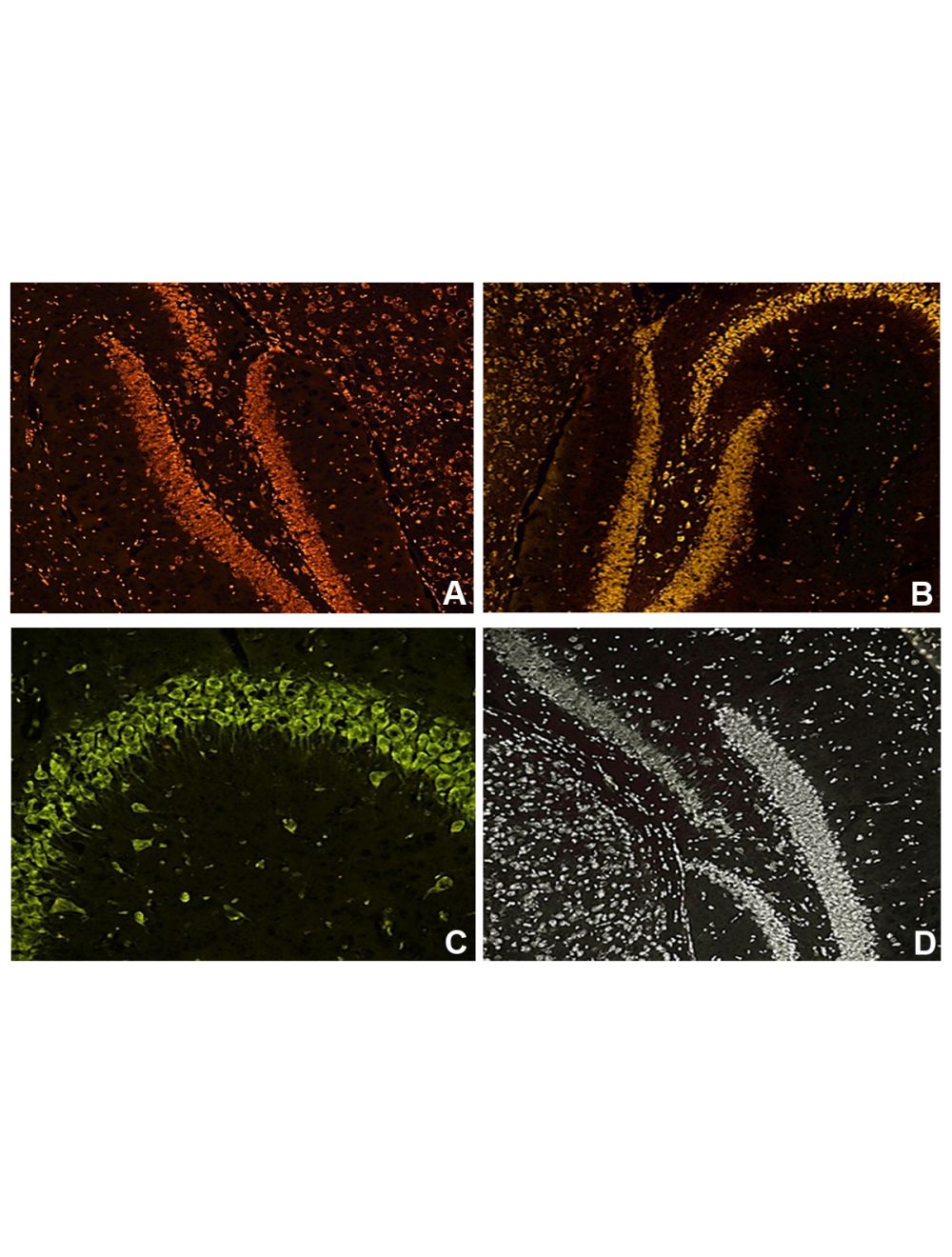

NisslFluor™ Nissl Sample Pack, Ready-to-Dilute™, Fluorescent Nissl Stains for Healthy Neurons
As low as
US$250.00
Only %1 left
Catalog Number
TR-SMP
- Product Name NisslFluor™ Nissl Sample Pack, Ready-to-Dilute™, Fluorescent Nissl Stains for Healthy Neurons
-
Product Description
The NisslFluor™ Nissl Sample Pack includes samples of NisslFluor™ Red (TR-200-NRD), Orange (TR-210-NOR), Green (TR-220-NGN), and Silver (TR-230-NSL). These products stain the soma and proximal parts of the dendrites, cytoplasm, and nuclei of neurons. Nissl substance is not usually present in the axon hillock or the axon itself. The NisslFluor™ fluorescent Nissl stains fluoresce in different colors and are excited by green (500-570 nm), blue (450-495 nm), or UV light (330-380 nm). These stains resist fading and provide high contrast, resolution, and brightness. Please refer to the individual Nissl stain inserts for complete instructions on each stain.
- Application(s) IF, ICC, IHC-Frozen, IHC-Paraffin-embedded
- Specificity Staining Nissl substances are present in neuronal soma and dendrites, not typically found in axons. Note: Like their bright-field counterparts, Nissl stains will stain all cells in the brain, not just the neurons. However, the staining of non-neuronal cells will be limited to their nuclei and a very thin rim of cytoplasm. By contrast, the cytoplasm and proximal dendrites of the larger neurons will be quite prominent and easily noticeable.
- Species Reactivity Human, Mouse, Other Mammals, Rat
- Concentration 10X
- Purity Description Thin layer chromatography using alumina plates and a solvent system of ethanol and water (3:1) revealed the presence of fluorescent isomers. No amount of starting material was detected.
- Regulatory Status For research use only.
Product Info
-
Product Description
The NisslFluor™ Nissl Sample Pack includes samples of NisslFluor™ Red (TR-200-NRD), Orange (TR-210-NOR), Green (TR-220-NGN), and Silver (TR-230-NSL). These products stain the soma and proximal parts of the dendrites, cytoplasm, and nuclei of neurons. Nissl substance is not usually present in the axon hillock or the axon itself. The NisslFluor™ fluorescent Nissl stains fluoresce in different colors and are excited by green (500-570 nm), blue (450-495 nm), or UV light (330-380 nm). These stains resist fading and provide high contrast, resolution, and brightness. Please refer to the individual Nissl stain inserts for complete instructions on each stain.
-
Related Products
Fluoro-Jade B (FJB) Powder for identifying Degenerating Neurons
Fluoro-Jade C (FJC) Powder for identifying Degenerating Neurons
NisslFluor™ Red, Ready-to-Dilute™, Fluorescent Nissl Stain for Healthy Neurons
NisslFluor™ Orange, Ready-to-Dilute™, Fluorescent Nissl Stain for Healthy Neurons
NisslFluor™ Green, Ready-to-Dilute™, Fluorescent Nissl Stain for Healthy Neurons
NisslFluor™ Silver, Ready-to-Dilute™, Fluorescent Nissl Stain for Healthy Neurons
- Application(s) IF, ICC, IHC-Frozen, IHC-Paraffin-embedded
- Application Details This multipack contains sample volumes of products TR-200-NRD, TR-210-NOR, TR-220-NGN, and TR-230-NSL. These products will stain the soma and proximal reaches of dendrites, cytoplasm, and nuclei of neurons a fluorescent red, orange, green, or sliver color depending upon the specific stain used and its illumination. Nissl substance is not generally found in the axon hillock or the axon itself. Please see our website and the individual NisslFluor™ Nissl stain product listing to download the associated protocol for complete use instructions. The number of slides stained will depend upon the species and size of the sections used and the staining container. Using a typical 50 mL Coplin jar, 160 slides (two mouse brain sections per slide) could easily be stained in one afternoon. The entire kit will stain at least 800 slides.
- Target Healthy neurons: Nuclei, Soma, and Dendrites.
- Specificity Staining Nissl substances are present in neuronal soma and dendrites, not typically found in axons. Note: Like their bright-field counterparts, Nissl stains will stain all cells in the brain, not just the neurons. However, the staining of non-neuronal cells will be limited to their nuclei and a very thin rim of cytoplasm. By contrast, the cytoplasm and proximal dendrites of the larger neurons will be quite prominent and easily noticeable.
- Target Host Species Human
- Species Reactivity Human, Mouse, Other Mammals, Rat
- Ex/Em Max Visualize sections of NisslFluor™ Red by green excitation using a TRITC-type filter block. Visualize sections of NisslFluor™ Orange by green excitation using a TRITC-type filter block. Visualize sections of NisslFluor™ Green by blue excitation using a FITC-type filter block. NisslFluor™ Silver RTD™ labeled neurons are visualized with UV light excitation (like those used for visualizing DAPI).
- Detection Method Fluorescence
-
Kit Components
Materials provided:
Four NisslFluor™ Stain Reagents: NisslFluor™ Red (TR-200-NRD), Orange (TR-210-NOR), Green (TR-220-NGN), and Silver (TR-230-NSL); 1 x 10 mL each; 10X (Dilute 1:10 prior to use). - Purity Description Thin layer chromatography using alumina plates and a solvent system of ethanol and water (3:1) revealed the presence of fluorescent isomers. No amount of starting material was detected.
- Format The reagents in the NisslFluor™ RTD™ (10X) are all supplied in a liquid format and are ready-to-dilute.
- Concentration 10X
- Reconstitution Instructions Dilute solutions as directed in the protocol instructions. Sometimes, small precipitates may be present in the stock or diluted solutions. Complete mixing of the diluted solutions usually dissolves the precipitates.
- Storage Instructions The stock solution can be stored for up to 6 months after receipt at 2-8ºC protected from light. No preservatives. Use sterile technique when handling and proper laboratory procedures. 1X solutions should be made fresh every day when in use.
- Storage Temperature 2-8ºC
- Batch Number Please see item label.
- Expiration Date Unopened kit 6 months at 2-8ºC protected from light. See Storage instructions for working solutions recommendations.
- Cellular Localization Nuclei, soma, dendrites
- Scientific Background Nissl substance, also termed chromophilic substance, exists as granular organelles in neuron cytoplasm. Named after its 19th-century discoverer, neurologist Franz Nissl, it consists of rough endoplasmic reticulum, RNA, and proteins and aids in protein synthesis for cell functions like growth and repair. Predominantly located in the neuron's soma, it's also in proximal dendrites but absent in axons. Its appearance and distribution vary based on the neuron type and activity. For instance, protein-synthesizing neurons like pyramidal cells contain large, basophilic Nissl chunks due to their maintenance needs, whereas sensory ganglion cells have dispersed, powdered forms. Typically, they're uniformly spread in the soma and dendritic regions, but their outward concentration might indicate neuronal damage or diseases, such as through central chromatolysis. Notably, after an axon is severed, its Nissl substance vanishes from the cell body. As a key neuronal activity and damage marker, changes in Nissl substance's size and distribution often signal stimuli response and are evident in conditions like Alzheimer's, where its reduction is linked to cognitive decline.
- Research Area Neuroscience
- Shipping Temperature Ambient
- UNSPSC CODE 60103920
- Regulatory Status For research use only.
