Application DetailsImmunocytochemistry (1:2,000 - 1:5,000) and Western Blot (1:1,000 - 1:5,000). Biosensis recommends optimal dilutions/concentrations should be determined by the end user.
TargetGreen fluorescent protein (GFP)
SpecificityGFP, cross reactivity with other mutant forms is expected as immunogen was a whole molecule GFP
Target Host SpeciesJellyfish
Species ReactivitySpecies Independent
Antibody HostRabbit
Antibody TypePolyclonal
Antibody IsotypeIgG
ConjugateUnconjugated
Immunogen DescriptionRecombinant AcGFP purified from E. coli
Purity DescriptionAffinity Purified
FormatLyophilized from PBS buffer pH 7.2-7.6 with 0.1% trehalose, and sodium azide
Reconstitution InstructionsSpin vial briefly before opening. Reconstitute with 100 µL sterile-filtered, ultrapure water. Centrifuge to remove any insoluble material.
Storage InstructionsStore lyophilized antibody at 2-8°C. After reconstitution divide into aliquots and store at -20°C for long-term storage. Store at 2-8°C short-term (up to 4 weeks). Avoid repetitive freeze/thaw cycles.
Batch NumberPlease see item label.
Expiration Date12 months after date of receipt (unopened vial).
Alternative NamesAequorea victoria green fluorescent protein
Scientific BackgroundThe green fluorescent protein (GFP) is a 27 kDa protein isolated originally from the jellyfish Aequoria victoria. It has an endogenous fluorochrome activity with excitation maximum at 395nm and emission maximum at 509 nm, which is similar to that of fluorescein. GFP can be expressed in fluorescent form in essentially any prokaryotic or eukaryotic cell. This GFP rabbit antibody was made against a recombinant GFP construct originating from an Aequoria species which was engineered to improve spectral properties and prevent oligomerization (1). This form of GFP, referred to as AcGFP, is 94% identical to the eGFP developed by Tsien and co-workers. The antibody can be used to verify the expression, size and stability of both AcGFP and eGFP fusion proteins in western blotting experiments and to amplify GFP signals in tissues of transgenic animals.
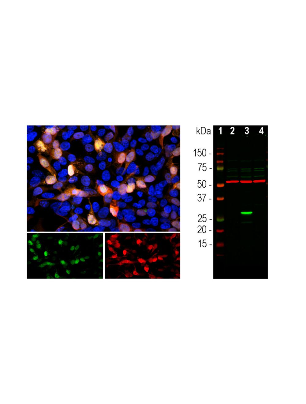
Left: Analysis of GFP expression (green) in transfected HEK293 cells by Immunocytochemistry. Cells were stained with rabbit anti-GFP antibody (red, 1:2,000). Blue: Hoechst staining for nuclei. Transfected cells appear orange-yellow due to the green (GFP) and red (anti-GFP antibody) color overlay. Right: Western blot analysis of GFP protein in transfected HEK293 lysate using rabbit antibody to GFP (green, 1:2,000). [1] protein standard, [2] non-transfected control cells, [3] transfected cells with GFP construct, [4] transfected cells with mCherry construct. A strong band at ~27 kDa corresponds to the expected molecular weight of GFP protein detected only in cells transfected with GFP construct. The antibody does not cross-react with mCherry protein. The same blot was simultaneously probed with a mouse antibody to beta-tubulin (red, ~ 50 kDa, lanes 2-4).
General ReferencesGurskaya NG et al. (2003) A colourless green fluorescent protein homologue from the non-fluorescent hydromedusa Aequorea coerulescens and its fluorescent mutants. Biochem J. 373(Pt 2):403-
 1800 605-5127
 +61 (0)8 8352 7711