Application DetailsWestern blotting (1:1,000-1:5,000) and Immunohistochemistry (1:2,000-1:5,000); Immunocytochemistry (1:1,000-1:5,000). Biosensis recommends optimal dilutions/concentrations should be determined by the end user.
Immunogen DescriptionC-terminal peptide of human IBA1 protein coupled to KLH.
Purity DescriptionWhole serum
FormatLyophilized with sodium azide.
Reconstitution InstructionsSpin vial briefly before opening. Reconstitute with 50 µL sterile-filtered, ultrapure water. Centrifuge to remove any insoluble material.
Storage InstructionsStore lyophilized antibody at 2-8°C. After reconstitution divide into aliquots and store at -20°C for long-term storage. Store at 2-8°C short-term (up to 4 weeks) with an appropriate antibacterial agent. Avoid repetitive freeze/thaw cycles.
Batch NumberPlease see item label.
Expiration Date12 months after date of receipt (unopened vial).
Alternative NamesAllograft inflammatory factor 1, AIF-1
Scientific BackgroundActin-binding protein that enhances membrane ruffling and RAC activation. Enhances the actin-bundling activity of LCP1. Binds calcium. Plays a role in RAC signaling and in phagocytosis. May play a role in macrophage activation and function. Promotes the proliferation of vascular smooth muscle cells and of T-lymphocytes. Enhances lymphocyte migration. Plays a role in vascular inflammation. Ref: uniprot.org
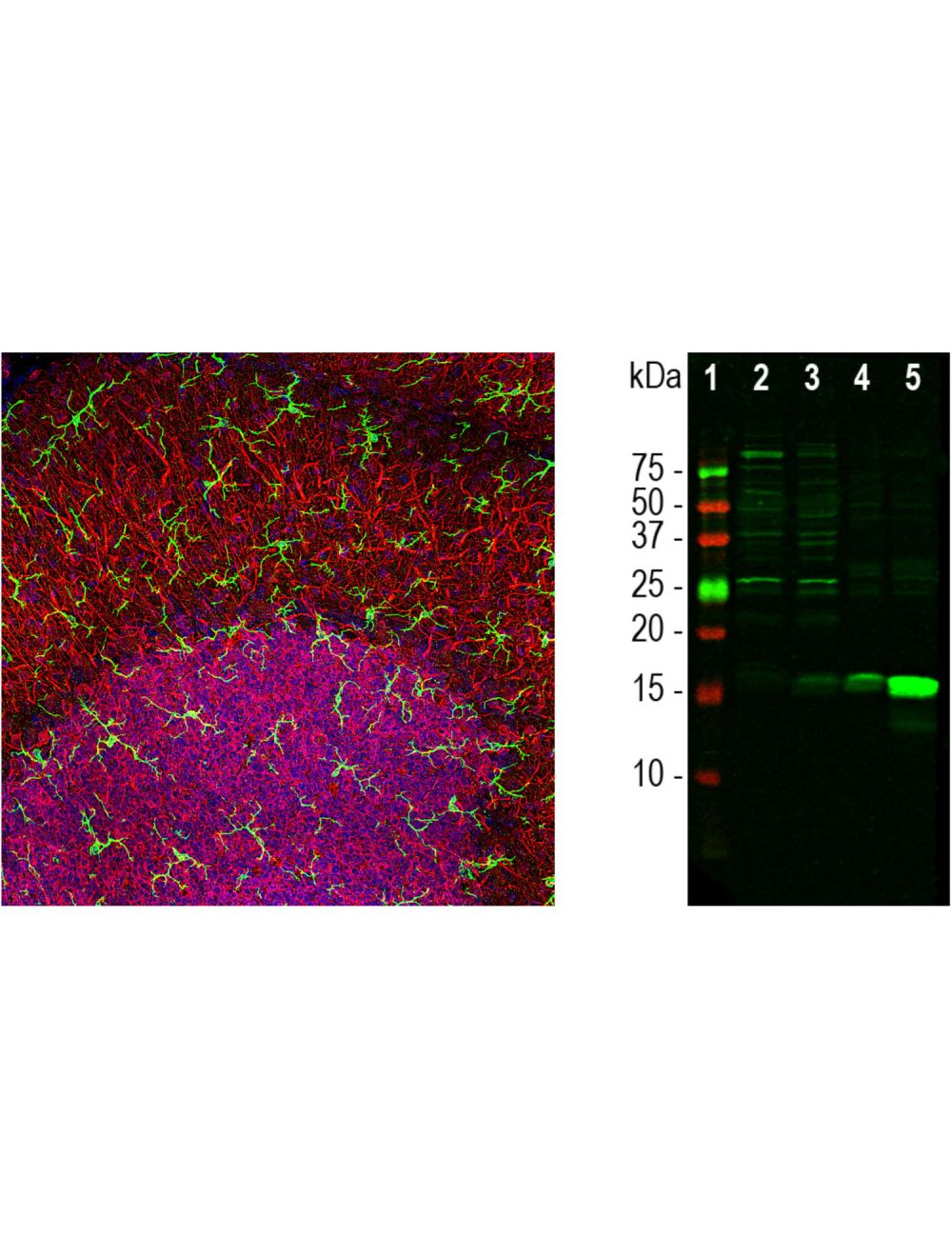
Left: Visualization of microglia (green) with anti-IBA antibody (1:1,000) by Immunohistochemistry. Image represents Z-stacked confocal image of rat cerebellar molecular layer at top and granular layer below. Microglia in their "surveilling" state demonstrate a small cell soma with fine, elongated processes spreading in three dimension. Red: MAP2-immunoreactivity in processes of Purkinje cells and the perikarya of granule cells, revealed with chicken anti-MAP2 antibody C-1382-50 (1:5,000). Blue: Nuclear DNA stained with DAPI. Right: Western blot analysis of tissue homogenates using rabbit anti-IBA1 antibody (1:1,000). Lane 1: Molecular weight standard; Lane 2: mouse brain; Lane 3: rat brain; Lane 4: mouse spleen; Lane 5: rat spleen. IBA1 appears at ~15-17 kDa. IBA1 is a relatively minor protein of brain and is much more abundant in spleen, as evidenced by different band intensities. The other bands seen in the CNS homogenates are of unknown origin, but do not appear to compromise the migroglia-specific staining seen with this antibody.
IBA1 staining in rat microglia (QBM Cell Science Catalog Number R-uG-535) by Immunocytochemistry. Primary antibody: 1:1,000 dilution. Magnification: 32X. Image courtesy of QBM Cell Science.
 1800 605-5127
 +61 (0)8 8352 7711