 1800 605-5127
 +61 (0)8 8352 7711
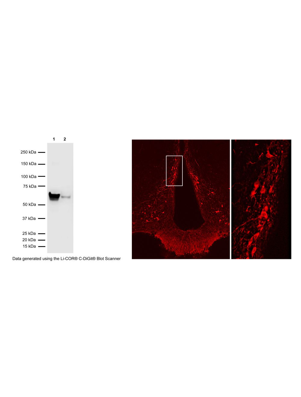

Tyrosine Hydroxylase (TH), Rabbit Polyclonal Antibody
- Product Name Tyrosine Hydroxylase (TH), Rabbit Polyclonal Antibody
- Product Description Rabbit anti-Tyrosine Hydroxylase (TH) Polyclonal Antibody (Unconjugated), suitable for IHC-Frozen.
- Alternative Names Tyrosine hydroxylase; Tyrosine 3-monooxygenase; L-tyrosine hydroxylase; Tyrosine 3-hydroxylase;
- Application(s) IHC-Frozen, WB
- Antibody Host Rabbit
- Antibody Type Polyclonal
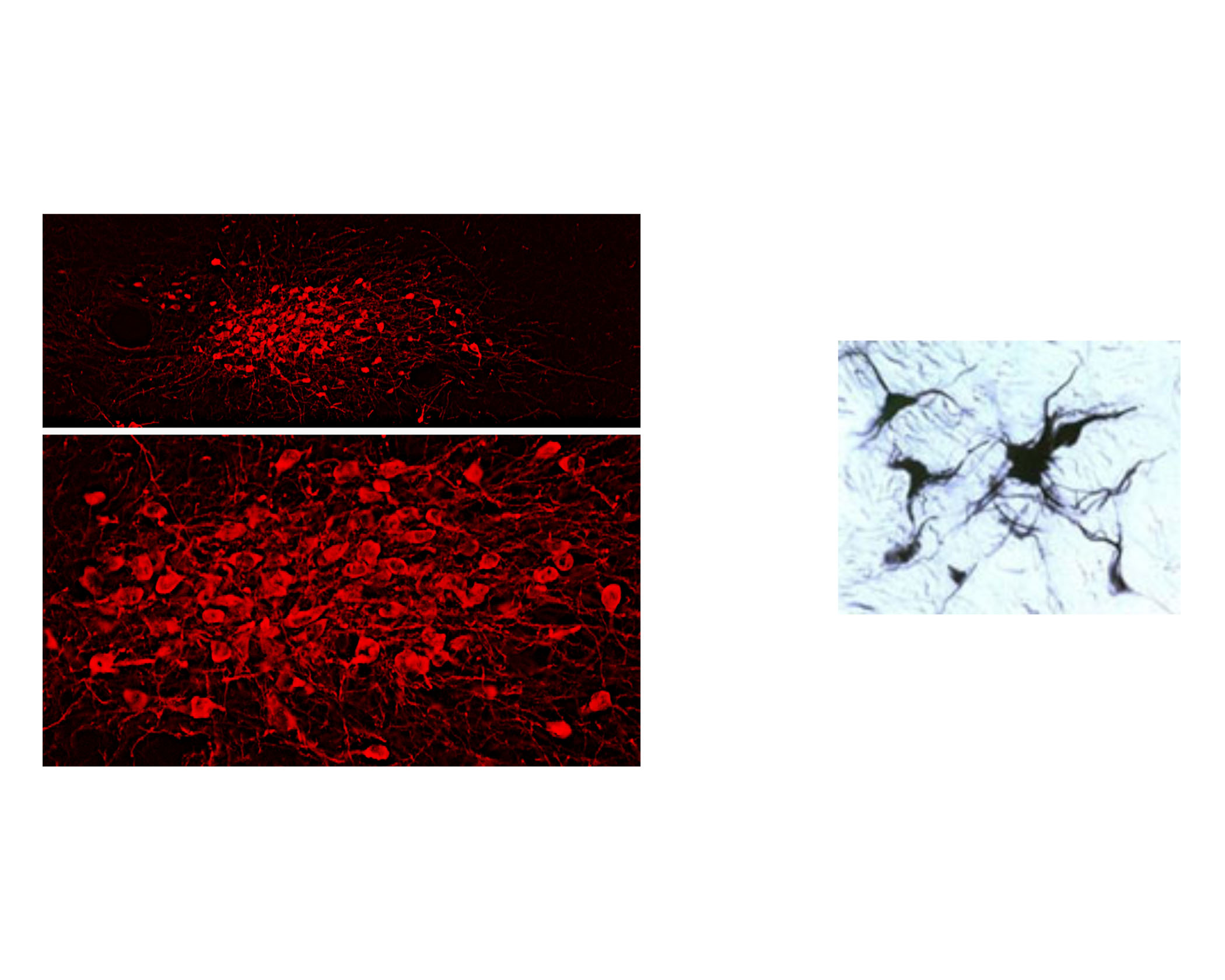
- Specificity IHC on brain shows a pattern of staining specific for TH containing neurons. This antibody is known to react with rat. Cross reactivity with other species has not yet been tested.
- Species Reactivity Human, Rat
- Immunogen Description A synthetic peptide (SPRFIGRRQSLIEDARK) as part of human Tyrosine Hydroxylase (aa: 32-47) conjugated to KLH
- Conjugate Unconjugated
- Purity Description Affinity purified
- Regulatory Status For research use only.
Product Info
- Product Description Rabbit anti-Tyrosine Hydroxylase (TH) Polyclonal Antibody (Unconjugated), suitable for IHC-Frozen.
-
Related Products
Tyrosine Hydroxylase (TH), Rabbit Polyclonal Antibody
- Application(s) IHC-Frozen, WB
-
Application Details
Immunohistochemistry (IHC): 0.5-1 ug/mL. This is a superb antibody for detection of tyrosine hydroxylase containing neurons exhibiting an intense labelling with a negligible background. This antiserum has proven extremely useful for staining of catecholaminergic neurons. It stains nicely and intensely dendritic processes and fine nerve terminals.
Western Blotting (WB): 0.5-2 ug/mL. This antibody demonstrates clear immunoreactivity for TH at 60 kDa in rat PC12 cell lysate and mouse brain homogenate. Biosensis recommends optimal dilutions/concentrations should be determined by the end user. - Target Tyrosine Hydroxylase (TH)
- Specificity IHC on brain shows a pattern of staining specific for TH containing neurons. This antibody is known to react with rat. Cross reactivity with other species has not yet been tested.
- Target Host Species Human
- Species Reactivity Human, Rat
- Antibody Host Rabbit
- Antibody Type Polyclonal
- Antibody Isotype IgG
- Conjugate Unconjugated
- Immunogen Description A synthetic peptide (SPRFIGRRQSLIEDARK) as part of human Tyrosine Hydroxylase (aa: 32-47) conjugated to KLH
- Purity Description Affinity purified
- Format Lyophilized
- Reconstitution Instructions Spin vial briefly before opening. Reconstitute in 50 µL sterile-filtered, ultrapure water. Centrifuge to remove any insoluble material.
- Storage Instructions After reconstitution keep aliquots at -20°C for a higher stability, and at 2-8°C with an appropriate antibacterial agent. Glycerol (1:1) may be added for an additional stability. Avoid repetitive freeze/thaw cycles.
- Batch Number Please see item label.
- Expiration Date 12 months after date of receipt (unopened vial).
- Alternative Names Tyrosine hydroxylase; Tyrosine 3-monooxygenase; L-tyrosine hydroxylase; Tyrosine 3-hydroxylase;
- Uniprot Number P04177
- Uniprot Number/Name P04177 (TY3H_RAT)
- Scientific Background Tyrosine hydroxylase (TH) is the rate-limiting enzyme in the synthesis of the catecholamines dopamine, epinephrine and norepinephrine. Therefore the regulation of the TH enzyme represents the central means for controlling the synthesis of these important catecholamines. FUNCTION: Plays an important role in the physiology of adrenergic neurons. CATALYTIC ACTIVITY: L-tyrosine + tetrahydrobiopterin + O2 = 3,4-dihydroxy-L-phenylalanine + 4a-hydroxytetrahydrobiopterin. COFACTOR: Fe(2+) ion. ENZYME REGULATION: Phosphorylation leads to an increase in the catalytic activity. PATHWAY: Catecholamine biosynthesis; first step. SUBUNIT: Homotetramer. PTM: In vitro, phosphorylation of Ser-19 increases the rate of Ser-40 phosphorylation, which results in enzyme opening and activation. SIMILARITY: Belongs to the biopterin-dependent aromatic amino acid hydroxylase family. The presence of different DNA sequences at the TH locus confers susceptibility to various disorders of the brain including manic-depression and schizophrenia. Parkinson's disease is also considered a TH deficiency as low dopamine levels are a consistent neurochemical abnormality.
- Shipping Temperature 25°C (ambient)
- UNSPSC CODE 41116161
- Regulatory Status For research use only.
Specifications

-
General References
Mallett, J. Trends in Pharmacological Science. 17(4): 129-135, 1996.
Haavik, J. et al. Mol. Neurobiology 16(3) :285-309, 199
Lewis DA, et al, Neuroscience 54: 477-, 1993
Kumer S.C. et al. Journal of Neurochemistry, 67(2) :443-462, 199
Haycock, J. Anal. Biochemistry 181: 259-266, 198
Haycock, J. Anal. Biochemistry 208: 397-399, 199
Renfroe, J.B., et al. Brain Res. Bull. 13: 109 - 126, 198
Xu, Z et a.l Neurosci. 82(3): 727, 1998