Product NameGrowth Associated Protein 43 (GAP-43), Rabbit Polyclonal Antibody
Product DescriptiongoogleRabbit anti-Growth Associated Protein 43 (GAP-43) Polyclonal Antibody (Unconjugated), suitable for WB, ICC.
Application(s)ICC, WB
Antibody HostRabbit
Antibody TypePolyclonal
SpecificityThe antibody reacts with a 43 kDa band by Western blot on bovine cerebellum homogenate. The molecular weight of the protein recognized can vary (~43-57 kDa) depending on the species and the percentage acrylamide used in the SDS-PAGE gel. It has also been used successfully for immunocytochemistry.
Species ReactivityBovine, Human, Mouse, Rat
Immunogen DescriptionC-terminal peptide 217-227 of rat and mouse GAP43, which is KEDPEADQEHA, to which an N terminal Cysteine residue was added to allow chemical coupling to Keyhole Limpet Hemocyanin carrier protein.
Product DescriptionRabbit anti-Growth Associated Protein 43 (GAP-43) Polyclonal Antibody (Unconjugated), suitable for WB, ICC.
Application(s)ICC, WB
Application DetailsWestern Blotting (WB). A dilution of 1:5,000 - 1:20,000 is recommended. A dilution of 1:500-2,000 is recommended for Immunocytochemistry (ICC). Biosensis recommends optimal dilutions/concentrations should be determined by the end user.
TargetGrowth Associated Protein 43 (GAP-43)
SpecificityThe antibody reacts with a 43 kDa band by Western blot on bovine cerebellum homogenate. The molecular weight of the protein recognized can vary (~43-57 kDa) depending on the species and the percentage acrylamide used in the SDS-PAGE gel. It has also been used successfully for immunocytochemistry.
Target Host SpeciesRat
Species ReactivityBovine, Human, Mouse, Rat
Antibody HostRabbit
Antibody TypePolyclonal
Antibody IsotypeIgG
ConjugateUnconjugated
Immunogen DescriptionC-terminal peptide 217-227 of rat and mouse GAP43, which is KEDPEADQEHA, to which an N terminal Cysteine residue was added to allow chemical coupling to Keyhole Limpet Hemocyanin carrier protein.
Purity DescriptionAffinity Purified
FormatLyophilized from PBS buffer pH 7.2-7.6 with 0.1% trehalose, and sodium azide
Reconstitution InstructionsSpin vial briefly before opening. Reconstitute with 100 µL sterile-filtered, ultrapure water. Centrifuge to remove any insoluble material.
Storage InstructionsStore lyophilized, unopened vial at 2-8°C or lower. After reconstitution, prepare aliquots and store at -20°C to -80°C for a higher stability. Avoid freeze-thaw cycles.
Batch NumberPlease see item label.
Expiration Date12 months after date of receipt (unopened vial).
Scientific BackgroundGAP43 is very abundant protein which is found concentrated in neurons. One group discovered it as one of three proteins which becomes unregulated during the regeneration of the toad optic nerve (1). Three GAPs (Growth associated proteins) were discovered, and the number 43 comes from the apparent SDS-PAGE molecular weight of the one named GAP43. The HGNC name for this protein is, not surprisingly, GAP43. Later work showed that GAP43 does not run on SDS-PAGE in a fashion which accurately reflects its molecular weight, and that GAP43 proteins from different species may run at different apparent molecular weights. Partly due to these features GAP43 were independently discovered by several different groups and therefore has several alternate names, such as protein F1, pp46, neuromodulin, neural phosphoprotein B-50 and calmodulin-binding protein P-57. In each case the number reflects the apparent SDS-PAGE molecular weight, and underlines the unusual properties of this molecule. Mammalian GAP43 proteins contains only 226-243 amino acids, and so the real molecular weight is 23.61-25.14 kDa. GAP43 has been extensively studied and is known to be a major protein kinase C substrate and to bind calmodulin avidly. GAP43 is anchored to the plasma membrane by palmitoylation modifications.
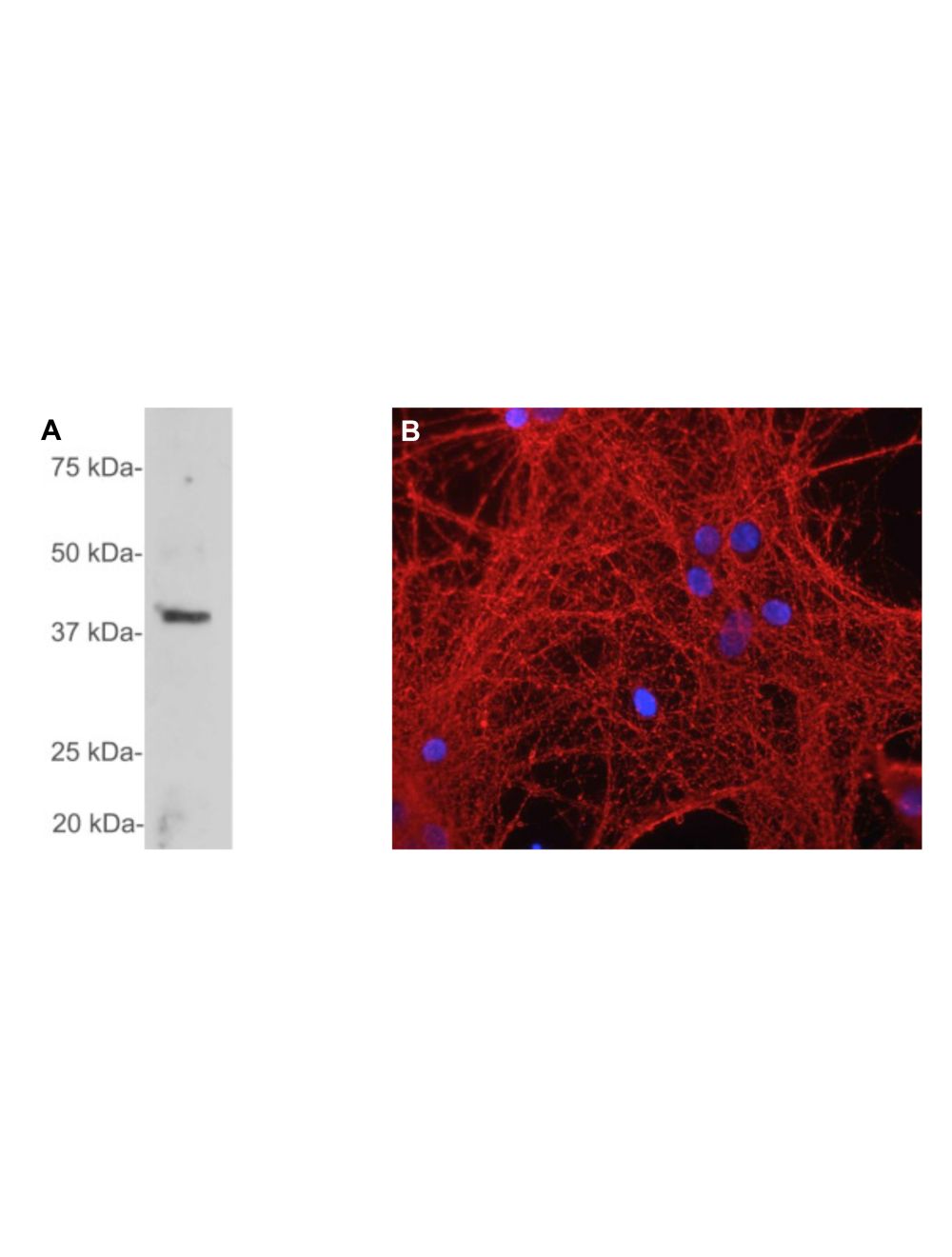
A: Western blot analysis of GAP43 expression in cow cerebellum. R-1651-100 detects a single band at ~43 kDa, which corresponds to full-length GAP43 protein. B: Immunofluorescence analysis of mixed neuron-glia cultures with GAP43 rabbit antibody R-1651-100 (red). DNA is stained blue.
Left: Analysis of cortical neuron-glial cell culture from E20 with rabbit antibody to GAP43 (green, 1:2,000) by Immunocytochemistry. Cells were co-stained with a mouse antibody to vimentin (red). Blue: DAPI nuclear stain. The GAP43 antibody labels protein expressed in the axonal membrane of neuronal cells, while the vimentin antibody stains intermediate filaments in fibroblasts and other non-neuronal cells. Right: Western blot analysis of tissue and cell lysates using rabbit antibody to GAP43 (green, 1:20,000). [1] protein standard, [2] rat brain, [3] rat spinal cord, [4] mouse brain, [5] mouse spinal cord, [6] SH-SY5Y cells, [7] C6. The single band at 43 kDa corresponds to GAP43 protein. The GAP43 protein is detected only in the lysates of neuronal origin. Rat glioblastoma C6 cells do not express GAP43 protein (lane 7).
General ReferencesSkene JH, Willard M. Changes in axonally transported proteins during axon regeneration in toad retinal ganglion cells. J. Cell Biol. 89:86-95 (1981).
 1800 605-5127
 +61 (0)8 8352 7711