 1800 605-5127
 +61 (0)8 8352 7711
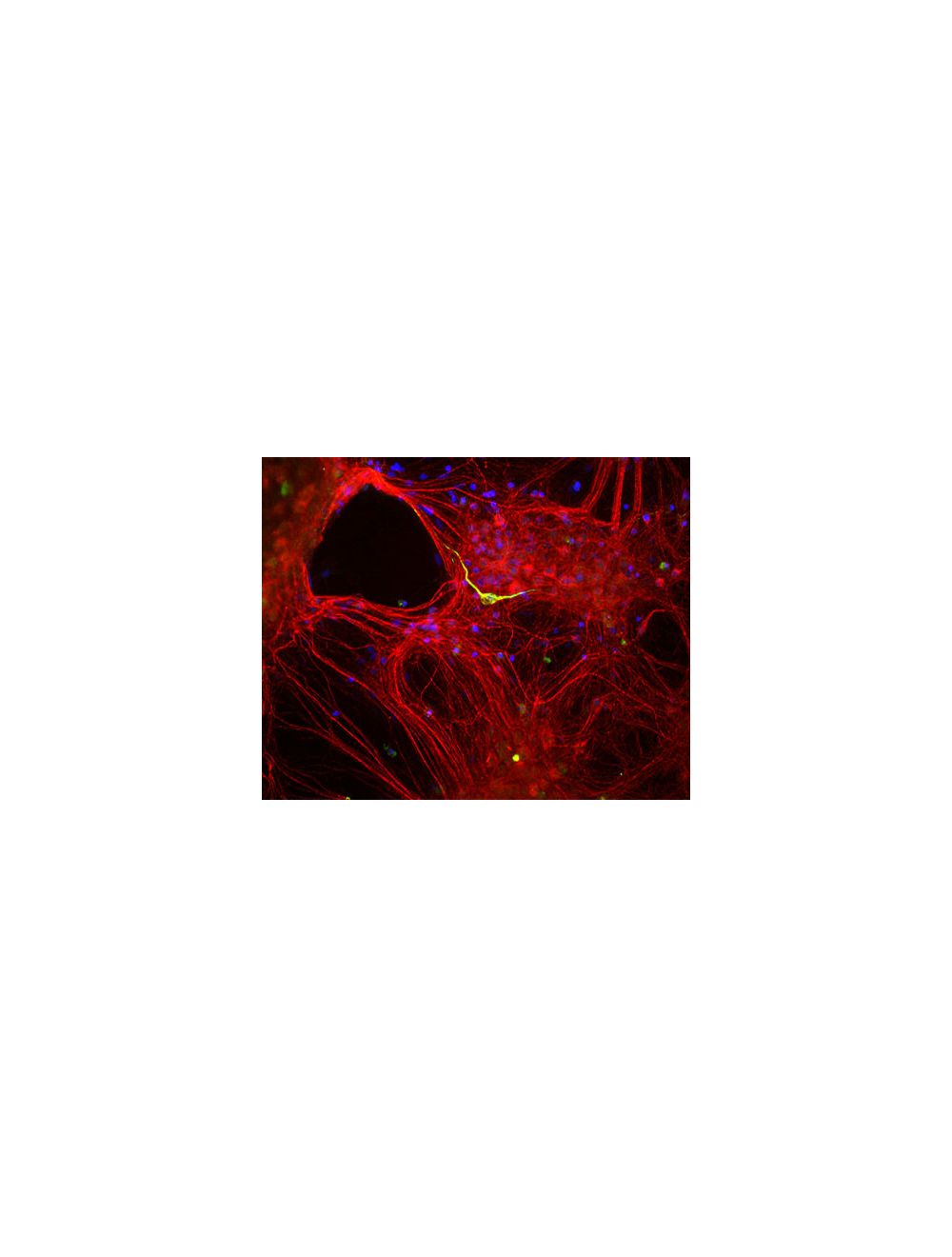

Internexin alpha (Alpha-Inx), Rabbit Polyclonal Antibody
- Product Name Internexin alpha (Alpha-Inx), Rabbit Polyclonal Antibody
- Product Description Rabbit anti-Internexin alpha (Alpha-Inx) Polyclonal Antibody (Unconjugated), suitable for WB, ICC, IHC-Frozen.
- Alternative Names 67 kDa neurofilament protein; NF-66; Neurofilament-66; Alpha-internexin; Alpha-Inx; Neurofilament 5; INA; NEF5;
- Application(s) ICC, IHC-Frozen, WB
- Antibody Host Rabbit
- Antibody Type Polyclonal
- Specificity The specificity of this antibody has been confirmed by WB. This antibody is specific for the 64-66 kDa alpha-internexin protein. Molecular weight will depend on species. Hu, Rat, Ms, Fel, and other mammals
- Species Reactivity Cat, Human, Mouse, Other Mammals, Rat
- Immunogen Description Recombinant alpha-internexin expressed and purified from E. coli
- Conjugate Unconjugated
- Purity Description Whole serum
- Regulatory Status For research use only.
Product Info
- Product Description Rabbit anti-Internexin alpha (Alpha-Inx) Polyclonal Antibody (Unconjugated), suitable for WB, ICC, IHC-Frozen.
- Application(s) ICC, IHC-Frozen, WB
- Application Details Western Blotting (WB), Immunocytochemistry (ICC) and Immunohistochemistry (IHC). A dilution of 1:10,000 - 1:20,000 is recommended for WB. A dilution of 1:500-1,000 is recommended for ICC and IHC. Biosensis recommends optimal dilutions/concentrations should be determined by the end user.
- Target Internexin alpha (Alpha-Inx)
- Specificity The specificity of this antibody has been confirmed by WB. This antibody is specific for the 64-66 kDa alpha-internexin protein. Molecular weight will depend on species. Hu, Rat, Ms, Fel, and other mammals
- Target Host Species Rat
- Species Reactivity Cat, Human, Mouse, Other Mammals, Rat
- Antibody Host Rabbit
- Antibody Type Polyclonal
- Antibody Isotype Mixed
- Conjugate Unconjugated
- Immunogen Description Recombinant alpha-internexin expressed and purified from E. coli
- Purity Description Whole serum
- Format Lyophilized with sodium azide.
- Reconstitution Instructions Spin vial briefly before opening. Reconstitute with 50 µL sterile-filtered, ultrapure water. Centrifuge to remove any insoluble material.
- Storage Instructions After reconstitution of lyophilized antibody, aliquot and store at -20°C for a higher stability. Avoid freeze-thaw cycles.
- Batch Number Please see item label.
- Expiration Date 12 months after date of receipt (unopened vial).
- Alternative Names 67 kDa neurofilament protein; NF-66; Neurofilament-66; Alpha-internexin; Alpha-Inx; Neurofilament 5; INA; NEF5;
- Uniprot Number P23565
- Uniprot Number/Name P23565 (AINX_RAT)
- Scientific Background Neurofilaments can be defined as the intermediate or 10nm diameter filaments found in neuronal cells. They are composed a mixture of subunits which often includes the neurofilament triplet proteins, NF-L, NF-M and NF-H. Neurofilaments may also include peripherin, alpha-internexin, nestin and in some cases vimentin. Alpha-internexin is a ~66 kDa Class IV intermediate filament subunit expressed in large amounts early in neuronal development, but is downregulated in many neurons as development procedes. Many classes of mature neurons contain alpha-internexin in addition to NF-L, NF-M and NF-H. In some mature neurons alpha-internexin is the only neurofilament subunit expressed. Antibodies to alpha-internexin are therefore unique probes to study and classify neuronal types and follow their processes in sections and in tissue culture. In addition the very early developmental expression of alpha-internexin means its presence is an early and convenient diagnostic feature of neuronal progenitors cells and other cell committed to the neuronal lineage.
- Shipping Temperature 25°C (ambient)
- UNSPSC CODE 41116161
- Regulatory Status For research use only.

