Product DescriptiongoogleRabbit anti-mCherry Polyclonal Antibody (Unconjugated), suitable for WB, ICC.
Application(s)ICC, WB
Antibody HostRabbit
Antibody TypePolyclonal
SpecificityThe antibody reacts with a band at ~28-30 kDa corresponding to intact full-length mCherry by Western blot on HEK293 cells transfected with mCherry vector. It has also been used successfully for immunocytochemistry.
Species ReactivitySpecies Independent
Immunogen DescriptionRecombinant full length mCherry.
Product DescriptionRabbit anti-mCherry Polyclonal Antibody (Unconjugated), suitable for WB, ICC.
Application(s)ICC, WB
Application DetailsWestern Blotting (WB) and Immunocytochemistry (IC). A dilution of 1:500 to 1:1,000 is recommended for WB. A dilution of 1:250 to 1:500 is recommended for IC. Biosensis recommends optimal dilutions/concentrations should be determined by the end user.
TargetmCherry
SpecificityThe antibody reacts with a band at ~28-30 kDa corresponding to intact full-length mCherry by Western blot on HEK293 cells transfected with mCherry vector. It has also been used successfully for immunocytochemistry.
Target Host SpeciesJellyfish
Species ReactivitySpecies Independent
Antibody HostRabbit
Antibody TypePolyclonal
Antibody IsotypeIgG
ConjugateUnconjugated
Immunogen DescriptionRecombinant full length mCherry.
Purity DescriptionAffinity purified
FormatLyophilized from PBS buffer pH 7.2-7.6 with 0.1% trehalose, and sodium azide
Reconstitution InstructionsSpin vial briefly before opening. Reconstitute with 100 µL sterile-filtered, ultrapure water. Centrifuge to remove any insoluble material.
Storage InstructionsStore lyophilized, unopened vial at 2-8°C or lower. After reconstitution, prepare aliquots and store at -20°C to -80°C for a higher stability. Avoid freeze-thaw cycles.
Batch NumberPlease see item label.
Expiration Date12 months after date of receipt (unopened vial).
Scientific BackgroundmCherry is an engineered derivative of one of a family of proteins originally isolated from Cnidarians (jelly fish, sea anemones and corals). The mCherry protein was derived from DsRed, a red fluorescent protein from so-called disc corals of the genus Discosoma. DsRed is a 223 amino acid ~28 kDa protein similar in size and properties to GFP, but, obviously, produces a red rather than a green fluorochrome. The original DsRed was engineered extensively in the Tsien lab to prevent it from forming tetramers and dimers and to modify and improve the spectral properties (1-3). The resulting monomeric protein is useful for applications such as Foerster Resonance Energy Transfer (FRET, also known as Fluorescence Resonance Energy Transfer). Several further cycles of mutation, directed modification and evolutionary selection produced mCherry, which is monomeric and has an excitation maximum at 587 nm and and emission maximum at 610 nm (4).
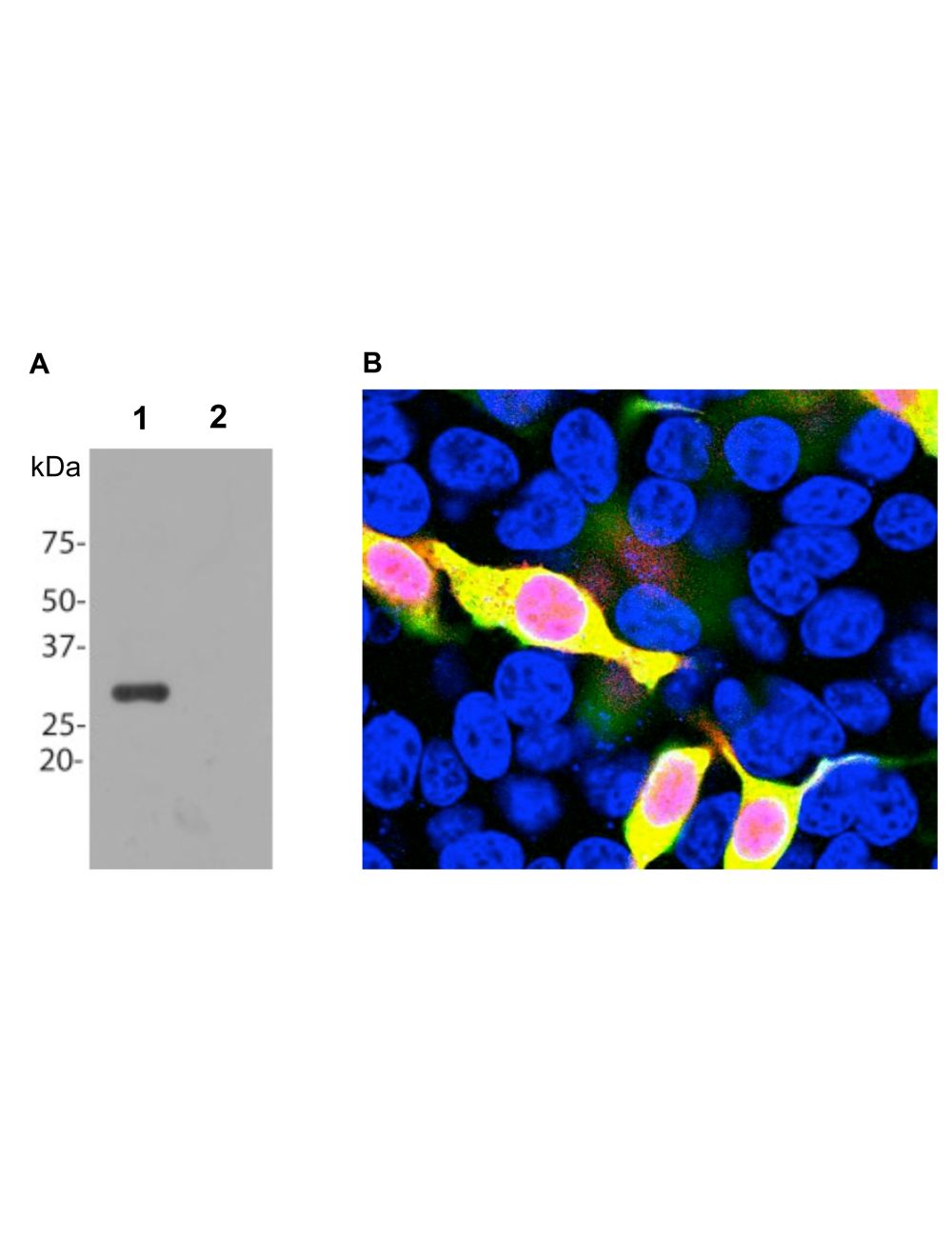
A: Western blot analysis of HEK293 cells transfected with pFin-EF1-mCherry vector. R-1654-100 reveals a strong protein band in transfected cells (Lane 1). Non-transfected control cells (Lane 2) show no mCherry protein band. B: Immunofluorescence analysis of mCherry-transfected HEK293 cells with R-1654-100. Specificity of R-1654-100 for mCherry is shown by superimposing mCherry fluorescence (red) with R-1654-100 antibody staining (green), which results in a yellow color. mCherry staining is also seen in the nucleus, which might be due to degradation of mCherry molecules, causing the release of the low molecular weight mCherry fluorochrome.
Western blot analysis of HEK293 cell lysates, and recombinant protein solutions using rabbit antibody to mCherry (green, 1:1,000). [1] protein standard, [2] HEK293, [3] HEK293 cells transfected with mCherry-HA construct, [4] mCherry recombinant protein, [5] GFP recombinant protein, and [6] HEK293 transfected with a full length GFP construct. A major band at about 28 kDa corresponds to mCherry protein. The antibody does not cross-react with GFP protein. The same blot was simultaneously probed with a chicken antibody to HSP60 (red, lanes 2-6).
General ReferencesBaird GS, Zacharias DA, Tsien RY. Biochemistry, mutagenesis, and oligomerization of DsRed, a red fluorescent protein from coral. Proc Natl Acad Sci U S A. 97:11984-9 (2000). Gross LA, Baird GS, Hoffman RC, Baldridge KK, Tsien RY. The structure of the chromophore within DsRed, a red fluorescent protein from coral. Proc Natl Acad Sci U S A. 97:11990-5 (2000). Heikal AA, Hess ST, Baird GS, Tsien RY, Webb WW. Molecular spectroscopy and dynamics of intrinsically fluorescent proteins: coral red (dsRed) and yellow (Citrine). Proc Natl Acad Sci U S A. 97:11996-2001 (2000). Shaner NC, Campbell RE, Steinbach PA, Giepmans BN, Palmer AE, Tsien RY. Improved monomeric red, orange and yellow fluorescent proteins derived from Discosoma sp. red fluorescent protein. Nature Biotechnology 22:1567-1572 (2004).
 1800 605-5127
 +61 (0)8 8352 7711