Product DescriptionRabbit anti-Vimentin Polyclonal Antibody (Unconjugated), suitable for WB, ICC.
Application(s)ICC, WB
Application DetailsWestern Blotting (WB) and Immunocytochemistry (ICC). A dilution of 1:5,000 - 1:10,000 is recommended for WB. A dilution of 1:1,000-5,000 is recommended for ICC. Biosensis recommends optimal dilutions/concentrations should be determined by the end user.
TargetVimentin
SpecificitySpecific for the ~55 kDa Vimentin protein.
Target Host SpeciesHuman
Species ReactivityHuman, Mouse, Other Mammals (Predicted)
Antibody HostRabbit
Antibody TypePolyclonal
Antibody IsotypeIgG
ConjugateUnconjugated
Immunogen DescriptionRecombinant human Vimentin purified from E. coli.
Purity DescriptionWhole serum
FormatLyophilized with sodium azide.
Reconstitution InstructionsSpin vial briefly before opening. Reconstitute with 100 µL sterile-filtered, ultrapure water. Centrifuge to remove any insoluble material.
Storage InstructionsAfter reconstitution of lyophilized antibody, aliquot and store at -20°C for a higher stability. Avoid freeze-thaw cycles.
Batch NumberPlease see item label.
Expiration Date12 months after date of receipt (unopened vial).
Scientific BackgroundVimentin is the major protein subunit of the 10nm or intermediate filaments protein found in many kinds of mesenchymal and epithelia cells. Vimentin is also found in many kinds of cells in tissue culture and in developing neuronal and astrocytic precursor cells in the central nervous system. Vimentin frequently forms copolymers with other intermediate filament proteins, such as GFAP (in many kinds of astrocytes), with desmin (in muscle cells) and neurofilament proteins (in developing neurons). Antibodies to vimentin are useful in studies of stem cells and generally to reveal the filamentous cytoskeleton.
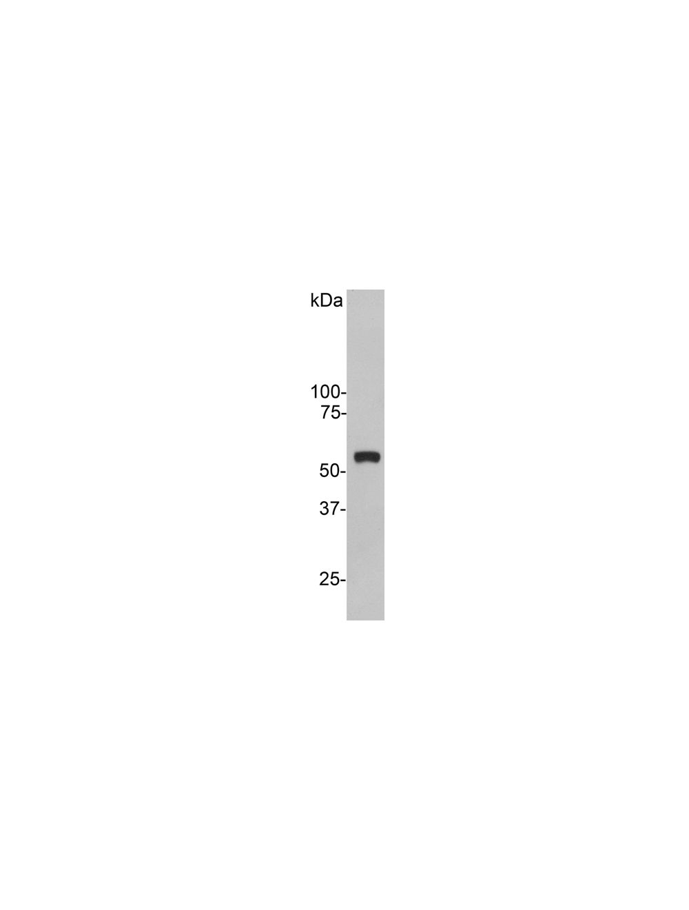
Western blot of crude extract of HeLa cells showing a single strong clean band at about 55 kDa.
HeLa cells stained with Rabbit anti-Vimentin (R-1699-100) (red) and counterstained with Monoclonal antibody to Lamin A/C (green, M-1689-100). DNA is blue. R-1699-100 antibody reveals strong cytoplasmic intermediate filament staining, while M-1689-100 antibody reveals strong nuclear lamina staining.
Left: Analysis of vimentin expression in HeLa cells by Immunocytochemistry with rabbit antibody to vimentin (green, 1:5,000). Cells were co-stained with an antibody to actin (red). Blue: DAPI nuclear stain. The vimentin antibody stains the 10 nm or intermediate filament network of the cytoskeleton. Right: Western blot analysis of whole cell lysates using rabbit antibody to vimentin (green, 1:5,000). [1] protein standard, [2] HeLa, [3] SH-SY5Y, [4] HEK293, [5] NIH-3T3. The strong band at around 55 kDa corresponds to vimentin protein.
 1800 605-5127
 +61 (0)8 8352 7711