SpecificityThe specificity of this antibody has been confirmed by WB. This antibody detects ~50 kDa Vimentin enzyme. Hu, Rat, Ms. It is predicted to react with other mammals due to sequence homology.
Species ReactivityHuman, Mouse, Other Mammals (Predicted), Rat
Immunogen DescriptionRecombinant human Vimentin purified from E.coli
Application DetailsWestern Blotting (WB) and Immunocytochemistry (ICC). A dilution of 1:5,000 - 1:10,000 is recommended for WB. A dilution of 1:1,000-5,000 is recommended for IC. Biosensis recommends optimal dilutions/concentrations should be determined by the end user.
TargetVimentin
SpecificityThe specificity of this antibody has been confirmed by WB. This antibody detects ~50 kDa Vimentin enzyme. Hu, Rat, Ms. It is predicted to react with other mammals due to sequence homology.
Target Host SpeciesHuman
Species ReactivityHuman, Mouse, Other Mammals (Predicted), Rat
Antibody HostChicken
Antibody TypePolyclonal
Antibody IsotypeIgY
ConjugateUnconjugated
Immunogen DescriptionRecombinant human Vimentin purified from E.coli
Purity DescriptionIgY
FormatLyophilized IgY preparation, with sodium azide.
Reconstitution InstructionsSpin vial briefly before opening. Reconstitute with 50 µL sterile-filtered, ultrapure water. Centrifuge to remove any insoluble material.
Storage InstructionsAfter reconstitution of lyophilized antibody, aliquot and store at -20°C for a higher stability. Avoid freeze-thaw cycles.
Batch NumberPlease see item label.
Expiration Date12 months after date of receipt (unopened vial).
Scientific BackgroundVimentins are class-III intermediate filaments specific to mesenchymal tissue. Vimentin is an important cytoskeletal component responsible for maintaining cell integrity and has a probable role in the intracellular transport of proteins such as lipoproteins between the nucleus and plasma membrane. Immunohistochemical staining for Vimentin is characteristic of sarcomas.
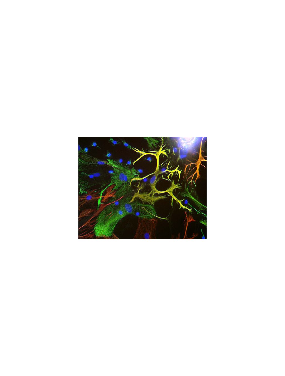
View of mixed neuron/glial cultures stained with Chicken polyclonal antibody to Vimentin C-1409-50 (green) and Rabbit polyclonal antibody to Glial Fibrillary Acidic Protein R-1374-50 (red). Vimentin is expressed alone in fibroblastic and endothelial cells, which are the flattened cells in the middle of the imate which appear green. Astrocytes may express primarily Glial Fibrillary Acidic Protein (GFAP), or GFAP and vimentin, and so appear red (GFAP only) or golden yellow (GFAP and Vimentin). In cells which express both GFAP and vimentin, the two protein assemble to produce heteropolymer filaments.
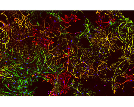
A view of neonatal rat brain cultures stained with Chicken polyclonal antibody to Vimentin C-1409-50 (red) and with Rabbit polyclonal antibody to GFAP R-1374-50 (green). These two proteins are found only in non-neuronal cells so you can\'t see any neurons, except for their nuclei in blue. Maturish astrocytes have only GFAP so appear green or have a mix of both proteins, so appear yellow. Some cells only have the vimentin (immature astrocytes, microglia and fibroblasts) and so appear red.
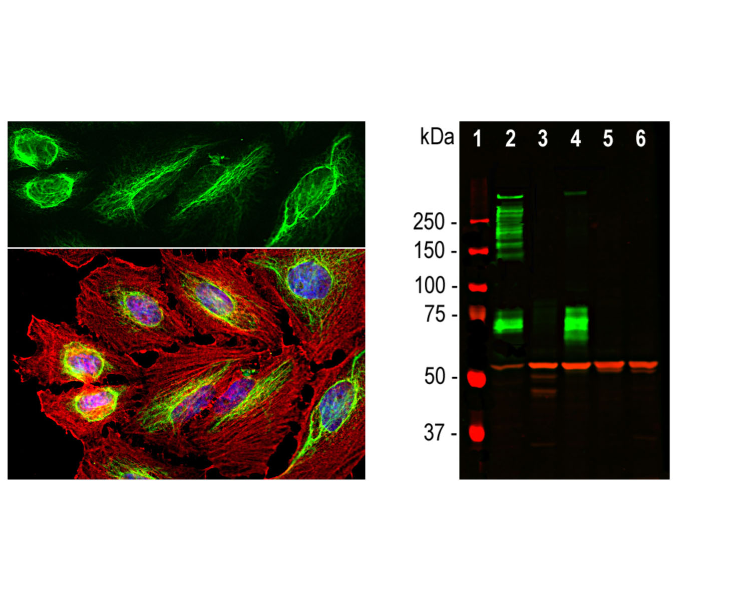
Left: Analysis of vimentin expression in HeLa cells by Immunocytochemistry. Cells were stained with chicken antibody to vimentin (1:10,000, green), and co-stained with a mouse anti-actin antibody (red). Blue: DAPI nuclear stain. The vimentin antibody stains the intermediate filament network, while the actin antibody labels the submembranous cytoskeleton, stress fibers, and bundles of actin associated with cell adhesion sites. Right: Western blot analysis of tissue and cell lysates using chicken antibody to Vimentin (1:5,000, red, lanes 2-6). [1] protein standard, [2] rat whole brain, [3] HeLa, [4] SH-SY5Y, [5] HEK293, [6] NIH-3T3. Vimentin protein appears as single band at around 50 kDa. The blot was simultaneously probed with a mouse antibody to MAP2C/D (green).
 1800 605-5127
 +61 (0)8 8352 7711