 1800 605-5127
 +61 (0)8 8352 7711
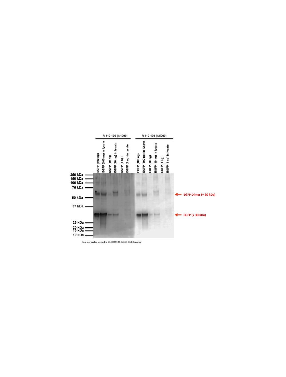

Enhanced green fluorescent protein (EGFP), Rabbit Polyclonal Antibody
Only %1 left
Catalog Number
R-110
Discontinued
- Product Name Enhanced green fluorescent protein (EGFP), Rabbit Polyclonal Antibody
- Product Description Rabbit anti-Enhanced green fluorescent protein (EGFP) Polyclonal Antibody (Unconjugated), suitable for WB, IHC-Frozen.
-
Replacement Product
Replaced by R-1838-100.
- Alternative Names Recombinant Enhanced Green Fluorescence Protein
- Application(s) IHC-Frozen, WB
- Antibody Host Rabbit
- Antibody Type Polyclonal
- Specificity This antibody is known to react with EGFP confirmed by IHC and WB.
- Species Reactivity Species Independent
- Immunogen Description Recombinant EGFP
- Conjugate Unconjugated
- Purity Description Whole serum
- Regulatory Status For research use only.
Product Info
- Product Description Rabbit anti-Enhanced green fluorescent protein (EGFP) Polyclonal Antibody (Unconjugated), suitable for WB, IHC-Frozen.
-
Replacement Product
Replaced by R-1838-100.
- Application(s) IHC-Frozen, WB
- Application Details IHC, Western blot. Recommended to be used at a dilution of 1:1000-1: 5000 for both applications. Biosensis recommends optimal dilutions/concentrations should be determined by the end user.
- Target Enhanced green fluorescent protein (EGFP)
- Specificity This antibody is known to react with EGFP confirmed by IHC and WB.
- Target Host Species Jellyfish
- Species Reactivity Species Independent
- Antibody Host Rabbit
- Antibody Type Polyclonal
- Antibody Isotype Mixed
- Conjugate Unconjugated
- Immunogen Description Recombinant EGFP
- Purity Description Whole serum
- Format Lyophilized
- Reconstitution Instructions Spin vial briefly before opening. Reconstitute in 100 µL sterile-filtered, ultrapure water. Centrifuge to remove any insoluble material.
- Storage Instructions After reconstitution keep aliquots at -20°C for a higher stability, and at 2-8°C with an appropriate antibacterial agent. Glycerol (1:1) may be added for an additional stability. Avoid repetitive freeze/thaw cycles.
- Batch Number Please see item label.
- Expiration Date 12 months after date of receipt (unopened vial).
- Alternative Names Recombinant Enhanced Green Fluorescence Protein
- Uniprot Number P42212
- Uniprot Number/Name P42212 (GFP_AEQVI)
- Scientific Background FUNCTION: Energy-transfer acceptor. Its role is to transduce the blue chemiluminescence of the protein aequorin into green fluorescent light by energy transfer. Fluoresces in vivo upon receiving energy from the Ca(2+)-activated photoprotein aequorin. BIOPHYSICOCHEMICAL PROPERTIES: Excitation max (nm): 488; Emission max (nm): 509; Extinction coefficient (Cm-1M-1): 61000. SUBUNIT: Monomer. TISSUE SPECIFICITY: Photocytes. PTM: Contains a chromophore consisting of modified amino acid residues. The chromophore is formed by autocatalytic backbone condensation between Xaa-N and Gly-(N+2), and oxidation of Tyr-(N+1) to didehydrotyrosine. Maturation of the chromophore requires nothing other than molecular oxygen. BIOTECHNOLOGY: Fluorescent proteins have become a useful and ubiquitous tool for making chimeric proteins, where they function as a fluorescent protein tag. Typically they tolerate N- and C-terminal fusion to a broad variety of proteins. They have been expressed in most known cell types and are used as a noninvasive fluorescent marker in living cells and organisms. They enable a wide range of applications where they have functioned as a cell lineage tracer, reporter of gene expression, or as a measure of protein-protein interactions. SIMILARITY: Belongs to the GFP family.
- Shipping Temperature 25°C (ambient)
- UNSPSC CODE 41116161
- Regulatory Status For research use only.
Specifications

-
General References
Chalfie, M., Tu, Y. et al (1994) Science 263, 802-805
Tsien, R. Y. (1998) Annu Rev Biochem 67, 509-544
Dardalhon, V. et al (1999) Hum Gene Ther 10, 5-14
Kain, S. R. (1999) DDT 4, 304-312