 1800 605-5127
 +61 (0)8 8352 7711
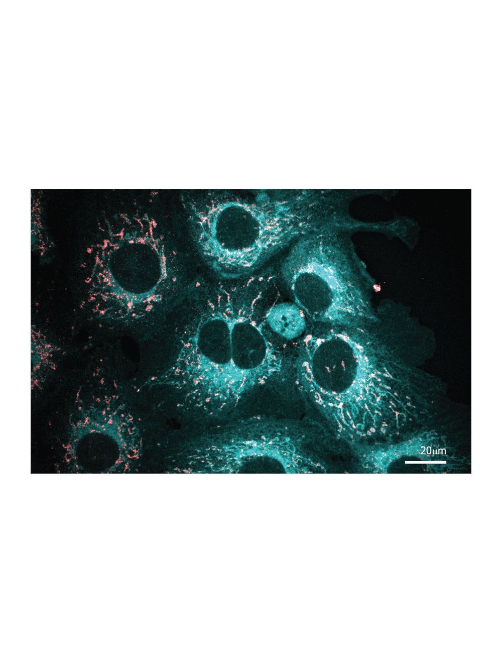

LipoFluor-ER2™ Ready-to-Dilute™, Endoplasmic Reticulum – Blue Tracing Reagent
As low as
US$297.00
Only %1 left
Catalog Number
TR-605
- Product Name LipoFluor-ER2™ Ready-to-Dilute™, Endoplasmic Reticulum – Blue Tracing Reagent
-
Product Description
LipoFluor-ER2™ is a cell-permeant stain for the endoplasmic reticulum with unique spectral properties, which makes it particularly useful for antibody double- and triple-labelling experiments. LipoFluor-ER2™ is highly compatible with green, red and far-red fluorophore-labelled antibodies and has been successfully used to image the endoplasmic reticulum in human and other mammalian cell lines. It passively diffuses across the plasma membrane into the cell, stains at low concentrations and has minimal cytotoxic effects. This tracing reagent can be used on live and fixed samples, is compatible with other dyes, shows excellent resistance to photobleaching and demonstrates low cytotoxicity.
- Application(s) IF, ICC, Live Cell IF
- Specificity Fluorescent imaging of the endoplasmic reticulum compartment. LipoFluor-ER2™ localises to the endoplasmic reticulum in a variety of live and fixed eukaryotic cells. It is highly cell permeable and produces fast results with ease and may be used in conjunction with antibody staining. The dye, however, cannot be fixed within the cells with post fixation methods.
- Species Reactivity Human, Mouse, Other Mammals, Rat
- Concentration 50mM
- Purity Description Thin layer chromatography using alumina plates and a solvent system of ethanol and water (3:1) revealed the presence of fluorescent isomers. No amount of starting material was detected.
- Regulatory Status For research use only.
Product Info
-
Product Description
LipoFluor-ER2™ is a cell-permeant stain for the endoplasmic reticulum with unique spectral properties, which makes it particularly useful for antibody double- and triple-labelling experiments. LipoFluor-ER2™ is highly compatible with green, red and far-red fluorophore-labelled antibodies and has been successfully used to image the endoplasmic reticulum in human and other mammalian cell lines. It passively diffuses across the plasma membrane into the cell, stains at low concentrations and has minimal cytotoxic effects. This tracing reagent can be used on live and fixed samples, is compatible with other dyes, shows excellent resistance to photobleaching and demonstrates low cytotoxicity.
-
Related Products
LipoFluor-P1™ Ready-to-Dilute™, Polar Lipid Tracing Reagent
LipoFluor-ER1™ Ready-to-Dilute™, Endoplasmic Reticulum Tracing Reagent
LipoFluor-MR™ Ready-to-Dilute™, Mitochondria Tracing Reagent
LipoFluor-P2™ Ready-to-Dilute™, Polar Lipid and Endoplasmic Reticulum Tracing Reagent
- Application(s) IF, ICC, Live Cell IF
-
Application Details
Fluorescent imaging of the endoplasmic reticulum compartment.
Sample types: live cells, fixed cells (4% paraformaldehyde)
Microscopy applications: epifluorescence, confocal, multicolour
Ex/Em: 405 nm / 500 nm; TR-605-ER2 can be excited by UV (~365 nm) or blue light (405 nm) sources with emissions collected using a wideband pass filter, or narrowband pass filter with an emission range 450-570 nm (Emmax = 500 nm). For confocal microscopy, the dye can be excited by a 400 nm steady state laser, and emission should be collected using a detector suited to blue fluorophores such as DAPI. Alternatively, a spectral detector set for the emission of ER-BTM stain (450-570 nm, Emmax = 500 nm) can be used.
Typical working concentration is 50µM (1:1000) dilution in appropriate buffer or cell culture medium. Biosensis recommends optimal dilutions/concentrations should be determined by the end user. - Target Endoplasmic reticulum
- Specificity Fluorescent imaging of the endoplasmic reticulum compartment. LipoFluor-ER2™ localises to the endoplasmic reticulum in a variety of live and fixed eukaryotic cells. It is highly cell permeable and produces fast results with ease and may be used in conjunction with antibody staining. The dye, however, cannot be fixed within the cells with post fixation methods.
- Target Host Species Human
- Species Reactivity Human, Mouse, Other Mammals, Rat
- Ex/Em Max Ex/Em: 405 nm / 570 nm
- Detection Method Fluorescence
-
Kit Components
Materials provided:
LipoFluor™ Stain Reagent, 1000X, 4 vials - Purity Description Thin layer chromatography using alumina plates and a solvent system of ethanol and water (3:1) revealed the presence of fluorescent isomers. No amount of starting material was detected.
- Format The reagents in the LipoFluor™ RTD™ (1000X) are all supplied in a liquid format and are ready-to-dilute.
- Concentration 50mM
- Reconstitution Instructions Dilute solutions as directed in the protocol instructions.
- Storage Instructions The liquid stock solution vials can be stored up to 12 months from date of receipt at 2-8°C, properly sealed and protected from light. Working solutions should be used within 24 hours and not stored for later use.
- Storage Temperature 2-8°C
- Batch Number Please see item label.
- Expiration Date If protected from light, the liquid 50 mM stock solution vials are stable for 12 months from date of receipt, at 2-8ºC. A vial should be diluted immediately prior to use and should be used within 24 hours for best results.
- Shipping Temperature 2-8°C (on cold packs)
- UNSPSC CODE 60103920
- Regulatory Status For research use only.
