 1800 605-5127
 +61 (0)8 8352 7711
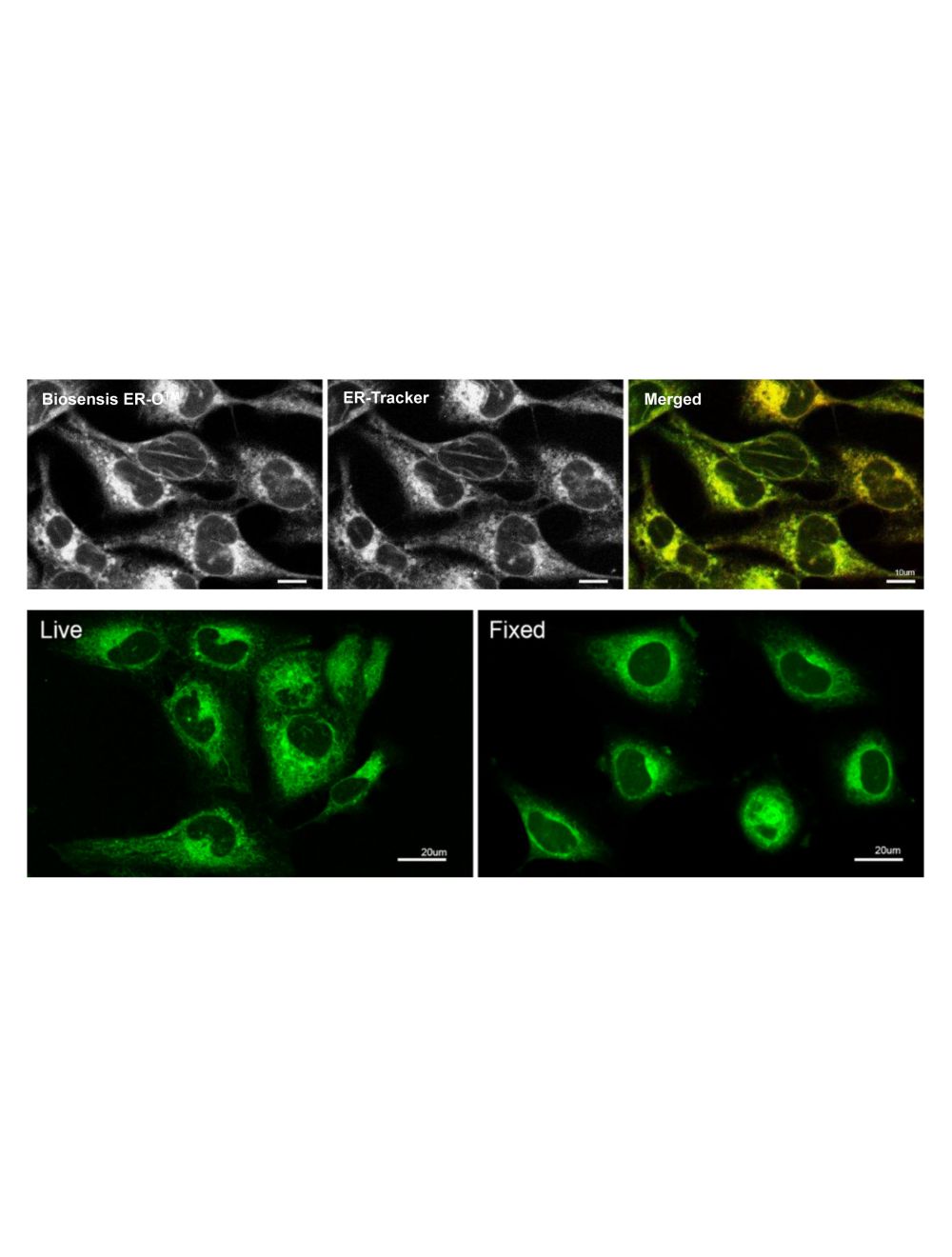

LipoFluor-ER1™ Ready-to-Dilute™, Endoplasmic Reticulum Tracing Reagent
As low as
US$297.00
Only %1 left
Catalog Number
TR-601
- Product Name LipoFluor-ER1™ Ready-to-Dilute™, Endoplasmic Reticulum Tracing Reagent
-
Product Description
LipoFluor-ER1™ is a cell-permeant stain for the endoplasmic reticulum. LipoFluor-ER1™ passively diffuses across the plasma membrane into the cell, stains at low concentrations and has minimal cytotoxic effects. It can be used as a real-time imaging reagent which can be imaged within minutes of addition and has minimal photobleaching. LipoFluor-ER1™ is easily washed from cells, and therefore is ideal for protocols which require intermittent monitoring of endoplasmic reticulum structures. This tracing reagent can be used on live and fixed samples and is compatible with other dyes. LipoFluor-ER1™ has a large Stokes Shift, shows excellent resistance to photobleaching and demonstrates low cytotoxicity.
- Application(s) IF, ICC, Live Cell IF
- Specificity Fluorescent imaging of the endoplasmic reticulum compartment. Cell penetration and localisation of LipoFluor-ER1™ has been confirmed in a range of cell lines, including prostate cells (PNT2, PNT1a, LNCaP, 22RV1 and DU145), cardiomyocytes (H9c2) and neuronal cells (PC-12).
- Species Reactivity Human, Mouse, Other Mammals, Rat
- Concentration 50mM
- Purity Description Thin layer chromatography using alumina plates and a solvent system of ethanol and water (3:1) revealed the presence of fluorescent isomers. No amount of starting material was detected.
- Regulatory Status For research use only.
Product Info
-
Product Description
LipoFluor-ER1™ is a cell-permeant stain for the endoplasmic reticulum. LipoFluor-ER1™ passively diffuses across the plasma membrane into the cell, stains at low concentrations and has minimal cytotoxic effects. It can be used as a real-time imaging reagent which can be imaged within minutes of addition and has minimal photobleaching. LipoFluor-ER1™ is easily washed from cells, and therefore is ideal for protocols which require intermittent monitoring of endoplasmic reticulum structures. This tracing reagent can be used on live and fixed samples and is compatible with other dyes. LipoFluor-ER1™ has a large Stokes Shift, shows excellent resistance to photobleaching and demonstrates low cytotoxicity.
-
Related Products
LipoFluor-P1™ Ready-to-Dilute™, Polar Lipid Tracing Reagent
LipoFluor-MR™ Ready-to-Dilute™, Mitochondria Tracing Reagent
LipoFluor-P2™ Ready-to-Dilute™, Polar Lipid and Endoplasmic Reticulum Tracing Reagent
LipoFluor-ER2™ Ready-to-Dilute™, Endoplasmic Reticulum – Blue Tracing Reagent
- Application(s) IF, ICC, Live Cell IF
-
Application Details
Fluorescent imaging of the endoplasmic reticulum compartment.
Sample types: live cells, fixed cells (4% paraformaldehyde), tissue (frozen only)
Epifluorescence microscopy: YES
Confocal microscopy: YES
Multiphoton imaging: YES
Infrared spectroscopy: NO
Raman spectroscopy: NO
Ex/Em: 405 nm / 570 nm.
The typical working concentration is 50µM (1:1000 dilution) in an appropriate buffer or cell culture medium. Biosensis recommends that the end user determine optimal dilutions/concentrations. - Target Endoplasmic reticulum
- Specificity Fluorescent imaging of the endoplasmic reticulum compartment. Cell penetration and localisation of LipoFluor-ER1™ has been confirmed in a range of cell lines, including prostate cells (PNT2, PNT1a, LNCaP, 22RV1 and DU145), cardiomyocytes (H9c2) and neuronal cells (PC-12).
- Target Host Species Human
- Species Reactivity Human, Mouse, Other Mammals, Rat
- Ex/Em Max Ex/Em: 405 nm / 570 nm
- Detection Method Fluorescence
-
Kit Components
Materials provided:
LipoFluor™ Stain Reagent, 1000X, 4 vials - Purity Description Thin layer chromatography using alumina plates and a solvent system of ethanol and water (3:1) revealed the presence of fluorescent isomers. No amount of starting material was detected.
- Format The reagents in the LipoFluor™ RTD™ (1000X) are all supplied in a liquid format and are ready-to-dilute.
- Concentration 50mM
- Reconstitution Instructions Dilute solutions as directed in the protocol instructions.
- Storage Instructions The liquid stock solution vials can be stored up to 12 months from date of receipt at 2-8°C, properly sealed and protected from light. Working solutions should be used within 24 hours and not stored for later use.
- Storage Temperature 2-8°C
- Batch Number Please see item label.
- Expiration Date If protected from light, the liquid 50 mM stock solution vials are stable for 12 months from date of receipt, at 2-8ºC. A vial should be diluted immediately prior to use and should be used within 24 hours for best results.
- Shipping Temperature 2-8°C (on cold packs)
- UNSPSC CODE 60103920
- Regulatory Status For research use only.

