Application DetailsWestern blotting (1:1,000) and Immunohistochemistry (frozen sections, 1:500-1:1,000). Biosensis recommends optimal dilutions/concentrations should be determined by the end user.
Product ValidationValidated by western blotting and immunohistochemical procedures.
TargetBeta-synuclein (β-synuclein, SNCB)
SpecificitySpecific for β-synuclein, does not cross-react with α- or γ-synuclein.
Target Host SpeciesHuman
Species ReactivityBovine, Human, Mouse, Pig, Rat
Antibody HostMouse
Antibody TypeMonoclonal
Antibody IsotypeIgG1
Clone Name6A10
ConjugateUnconjugated
Immunogen DescriptionC-terminal peptide of human β-synuclein (EPEGESYEDPPQEEYQEYEPEA) coupled to KLH.
Immunogen Length22 amino acids.
SequenceEPEGESYEDPPQEEYQEYEPEA
EpitopeC-terminus.
Purity DescriptionProtein G purified
Physical StateSolid.
FormatLyophilized from PBS buffer pH 7.2-7.6 with 0.1% trehalose, and sodium azide
Reconstitution InstructionsSpin vial briefly before opening. Reconstitute with 100 µL sterile-filtered, ultrapure water to achieve a 1 mg/mL concentration. Centrifuge to remove any insoluble material.
Storage InstructionsStore lyophilized antibody at 2-8°C. After reconstitution divide into aliquots and store at -20°C for long-term storage. Store at 2-8°C short-term (up to 4 weeks). Avoid repetitive freeze/thaw cycles.
Batch NumberPlease see item label.
Expiration Date12 months after date of receipt (unopened vial).
Scientific BackgroundNon-amyloid component of senile plaques found in Alzheimer disease. Could act as a regulator of SNCA aggregation process. Protects neurons from staurosporine and 6-hydroxy dopamine (6OHDA)-stimulated caspase activation in a p53/TP53-dependent manner. Contributes to restore the SNCA anti-apoptotic function abolished by 6OHDA. Not found in the Lewy bodies associated with Parkinson disease (Ref: uniprot.org).
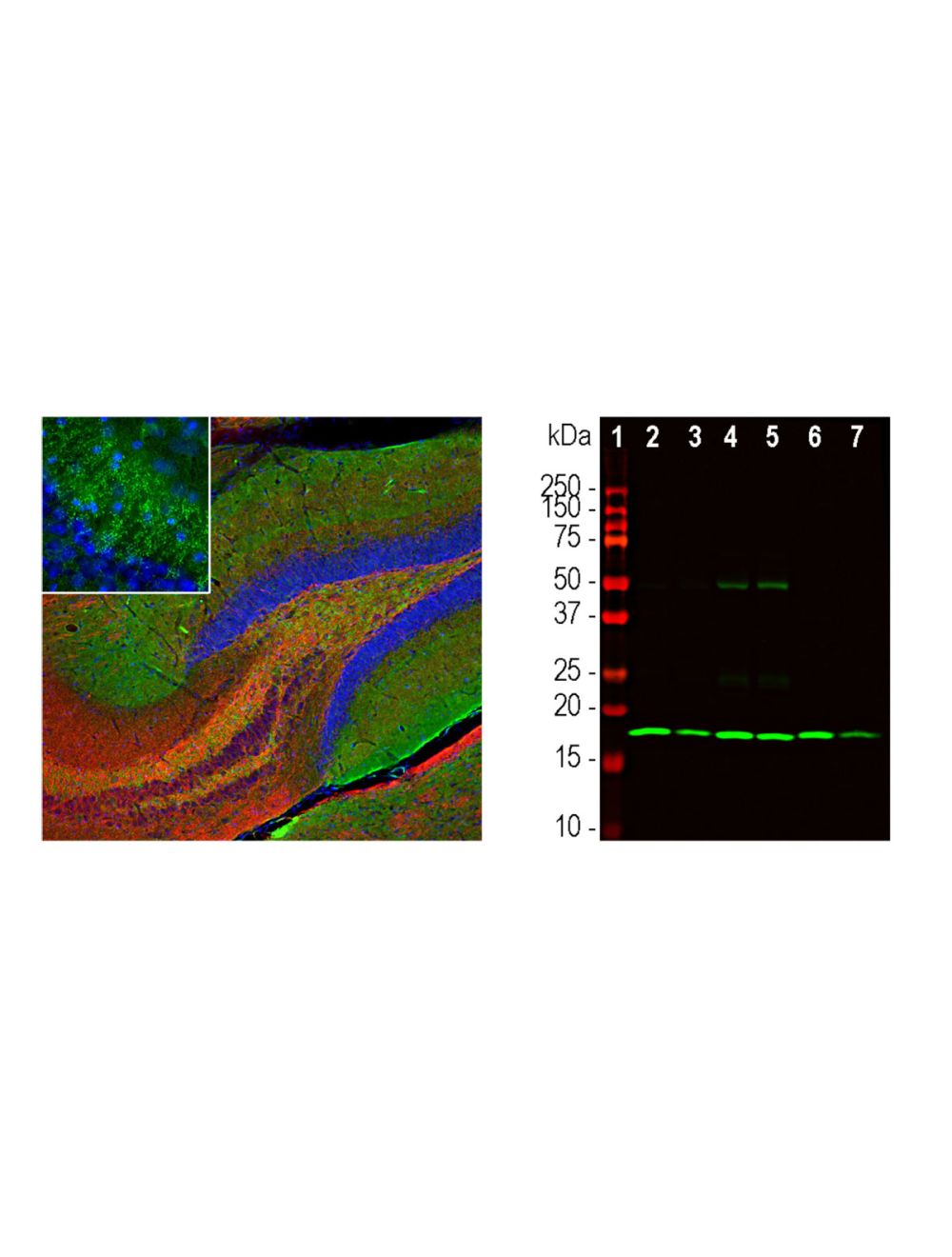
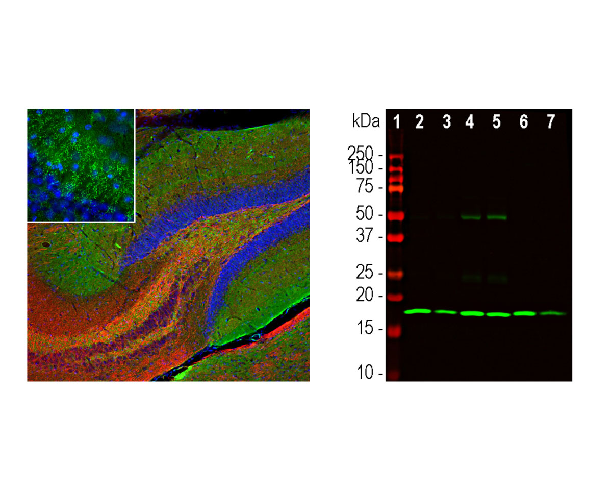
Left: Analysis of beta-synuclein expression in mouse hippocampus section by Immunohistochemistry. Beta-synuclein (green) was detected with mouse antibody to beta-synuclein (1:500), and sections co-stained with a rabbit antibody to neurofilament light (NF-L, C-terminal, red). Blue: Hoechst nuclear stain. IHC Method: Following transcardial perfusion of mouse with 4% paraformaldehyde, brain was post-fixed for 24 hours, cut to 45 μm, and free-floating sections were stained with above antibodies. The beta-synuclein antibody detects protein concentrated in synaptic regions, while the NF-L antibody labels dendrites and axons of neuronal cells. Right: Western blot analysis of tissue homogenates using mouse antibody to beta-synuclein (green, 1:1,000). Lane 1: molecular weight marker; Lane 2: rat cortex; Lane 3: rat cerebellum; Lane 4: mouse cortex; Lane 5: mouse cerebellum; Lane 6: cow cortex; Lane 7: cow cerebellum. A strong band at about 17 kDa corresponds to beta-synuclein protein.
 1800 605-5127
 +61 (0)8 8352 7711